Clinicians looking at an x-ray on a screen

Pleural Disease Full Guideline

BTS Guideline on Pleural Disease

Cite this article as: Evans HJ, Gibson NA, Bennett J. On behalf of the BTS Pleural Guideline Development Group, et al British Thoracic Society Guideline for pleural disease. Thorax 2023;78:s1-s42.

Pleural disease is common and represents a major and rapidly developing subspecialty that presents to many different hospital services. Since the last British Thoracic Society (BTS) Guideline for pleural disease published in 2010,(3-9) many high-quality and practice changing studies, using patient-centred outcomes, have been published. The paradigms for the investigation and management of pleural disease have therefore shifted. For example, ambulatory treatments have become much more prominent in the management of pleural disease. This guideline aims to capture this evidence and use it to answer the most important questions relevant to today’s practice.

This Guideline provides advice on the investigation and management of:

i. Spontaneous pneumothorax 
ii. Undiagnosed unilateral pleural effusion
iii. Pleural infection
iv. Pleural malignancy

Please note that this has also been published in conjunction with the corresponding BTS Clinical Statement on Pleural Procedures. Both pieces of guidance are contained within the Full Supplement document found below.

The Guideline and Summary can also be accessed via the Thorax journal here: Full Guideline and Summary

Watch a BTS webinar introducing the new Pleural Guideline and Clinical Statement.

Recommendations

i- Spontaneous Pneumothorax

Acute management for spontaneous pneumothorax 

Recommendations

  • Conservative management can be considered for the treatment of minimally symptomatic (ie, no significant pain or breathlessness and no physiological compromise) or asymptomatic primary spontaneous pneumothorax in adults regardless of size. (Conditional—by consensus)

  • Ambulatory management should be considered for the initial treatment of primary spontaneous pneumothorax (PSP) in adults with good support, and in centres with available expertise and follow-up facilities. (Conditional)

  • In patients not deemed suitable for conservative or ambulatory management, needle aspiration or tube drainage should be considered for the initial treatment of primary spontaneous pneumothorax in adults. (Conditional)

  • Chemical pleurodesis can be considered for the prevention of recurrent of secondary spontaneous pneumothorax (SSP) in adults (eg, patients with severe chronic obstructive pulmonary disease who significantly decompensated in the presence of a pneumothorax, even during/after the first episode). (Conditional)

  • Thoracic surgery can be considered for the treatment of pneumothorax in adults at initial presentation if recurrence prevention is deemed important (eg, patients presenting with tension pneumothorax, or those in high-risk occupations). (Conditional)

Good practice points

  • When establishing local ambulatory treatment pathways, planning and coordination between with the emergency department, general medicine and respiratory medicine is vital.

  • When performing chemical pleurodesis for the treatment of pneumothorax in adults, adequate analgesia should be provided before and after treatment.

  • All treatment options should be discussed with the patient to determine their main priority, with consideration for the least invasive option.

Optimal management after the resolution of a first episode of pneumothorax

Good practice points

  • Elective surgery may be considered for patients in whom recurrence prevention is deemed important (eg, at-risk professionals (divers, airline pilots, military personnel), or those who developed a tension pneumothorax at first episode).

  • Elective surgery should be considered for patients with a second ipsilateral or first contralateral pneumothorax.

  • Discharge and activity advice should be given to all patients post pneumothorax.

Optimal management for spontaneous pneumothorax and ongoing air leak

Good practice points

  • If a patient is not considered fit for surgery, autologous blood pleurodesis or endobronchial therapies should be considered for the treatment of pneumothorax with persistent air leak in adults.

Optimal surgical approach and surgical operation for pneumothorax management

Recommendations

  • Video-assisted thoracoscopy access can be considered for surgical pleurodesis in the general management of pneumothorax in adults. (Conditional)

  • Thoracotomy access and surgical pleurodesis should be considered for the lowest level of recurrence risk required for specific (eg, high-risk) occupations. (Conditional)

  • Surgical pleurodesis and/or bullectomy should be considered for the treatment of spontaneous pneumothorax in adults. (Conditional)

ii- Investigation of the undiagnosed unilateral pleural effusion

Radiology for diagnosing unilateral pleural effusions of benign aetiology

Good practice points

  • Imaging findings of a unilateral pleural effusion should be interpreted in the context of clinical history and knowledge of pleural fluid characteristics.

  • CT follow-up should be considered for patients presenting with pleural infection to exclude occult malignancy if there are ongoing symptoms, or other clinically concerning features.

  • Positron emission tomography-CT (PET-CT) should not be used in the assessment of pleural infection.

Image-guided versus non-image-guided intervention for suspected unilateral pleural effusion

Recommendation

  • Image-guided thoracentesis should always be used to reduce the risk of complications. (Strong—by consensus)

Optimal volume and container for pleural aspiration samples

Recommendations

  • 25–50 mL of pleural fluid should be submitted for cytological analysis in patients with suspected malignant pleural effusion (MPE). (Strong—by consensus)

  • Pleural fluid should be sent in both plain and blood culture bottle tubes in patients with suspected pleural infection. (Strong—by consensus)

Good practice points

  • At least 25 mL, and where possible 50 mL, of pleural fluid should be sent for initial cytological examination.

  • If volumes of ≥25 mL cannot be achieved, smaller volumes should be sent, but clinicians should be aware of the reduced sensitivity.

  • If small volume aspirate (<25 mL) has been non-diagnostic, a larger volume should be sent, if achievable, except when there is high suspicion of a tumour type associated with low pleural fluid cytology sensitivity (especially mesothelioma).

  • Pleural fluid samples should be processed by direct smear and cell block preparation.

  • In patients with an undiagnosed pleural effusion where pleural infection is possible and volume of fluid sample available allows, microbiological samples should be sent in both white top containers and volumes of 5–10 mL inoculated into (aerobic and anaerobic) blood culture bottles.

  • In cases where volume available does not allow 5–10 mL inoculation, volumes of 2–5 mL should be prioritised to blood culture bottles rather than a plain, sterile container.

Pleural fluid tests (biomarkers) for diagnosing unilateral pleural effusion

Recommendations

  • Pleural fluid cytology should be used as an initial diagnostic test in patients with suspected secondary pleural malignancy, accepting that a negative cytology should lead to consideration of further investigation. (Conditional)

  • Pleural fluid biomarkers should not be used for diagnosing secondary pleural malignancy. (Conditional)

  • In high prevalence populations, pleural fluid adenosine deaminase (ADA) and/or interferon gamma (IFN-gamma) test(s) can be considered for diagnosing tuberculous pleural effusion. (Conditional)

  • In low prevalence populations, pleural fluid ADA can be considered as an exclusion test for tuberculous pleural effusion. (Conditional)

  • Tissue sampling for culture and sensitivity should be the preferred option for all patients with suspected tuberculous pleural effusion. (Strong—by consensus)

  • Pleural fluid antinuclear antibody (ANA) should be considered to support a diagnosis of lupus pleuritis. (Conditional)

Good practice points

  • The clinical utility of pleural fluid cytology varies by tumour subtype, including diagnostic sensitivity and predictive value for response to subsequent cancer therapies. This should be taken into consideration when planning the most suitable diagnostic strategy (eg, direct biopsies in those with a likely low cytological yield can be considered).

  • Pleural fluid N-terminal prohormone brain natriuretic peptide (NT-proBNP) is useful when considering heart failure as a cause in unilateral pleural effusions but not superior to serum NT-proBNP and therefore should not be ordered routinely.
Serum biomarkers for diagnosing unilateral pleural effusion

Recommendation

  • Serum NT-proBNP should be considered to support a diagnosis of heart failure in patients with unilateral pleural effusion suspected of having heart failure. (Conditional)

Good practice points

  • Serum biomarkers should not currently be used to diagnose secondary pleural malignancy, pleural infection or autoimmune pleuritis.

  • Serum biomarkers should not routinely be used to diagnose tuberculous pleural effusion, but may be considered in high prevalence areas.

  • Serum biomarkers, including NT-proBNP, should not be used in isolation for diagnosing unilateral pleural effusion, as multiple conditions may co-exist.

Pleural biopsy for diagnosing unilateral pleural effusion

Recommendations

  • Thoracoscopic or image-guided pleural biopsy may be used depending on the clinical indication and local availability of techniques (including need for control of pleural fluid). (Strong)

  • Blind (non-image-guided) pleural biopsies should not be conducted. (Strong—by consensus)

iii- Pleural Infection

Predicting clinical outcomes of pleural infection

Recommendation

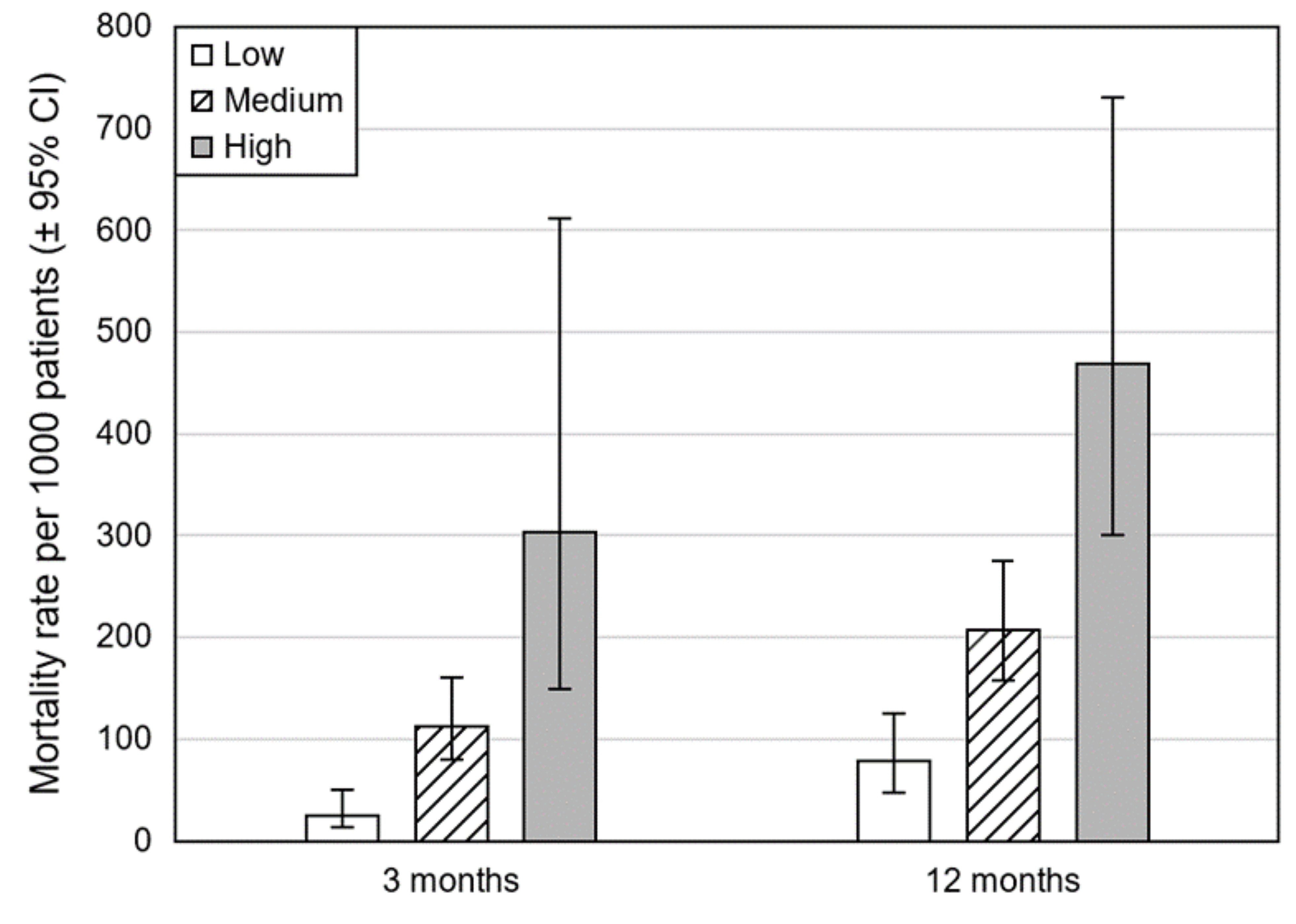
  • Renal, age, purulence, infection source, dietary factors (RAPID) scoring should be considered for risk stratifying adults with pleural infection and can be used to inform discussions with patients regarding potential outcome from infection. (Conditional)

Pleural fluid, or radiology parameters for determining which patients can be treated with intercostal drainage

Recommendations

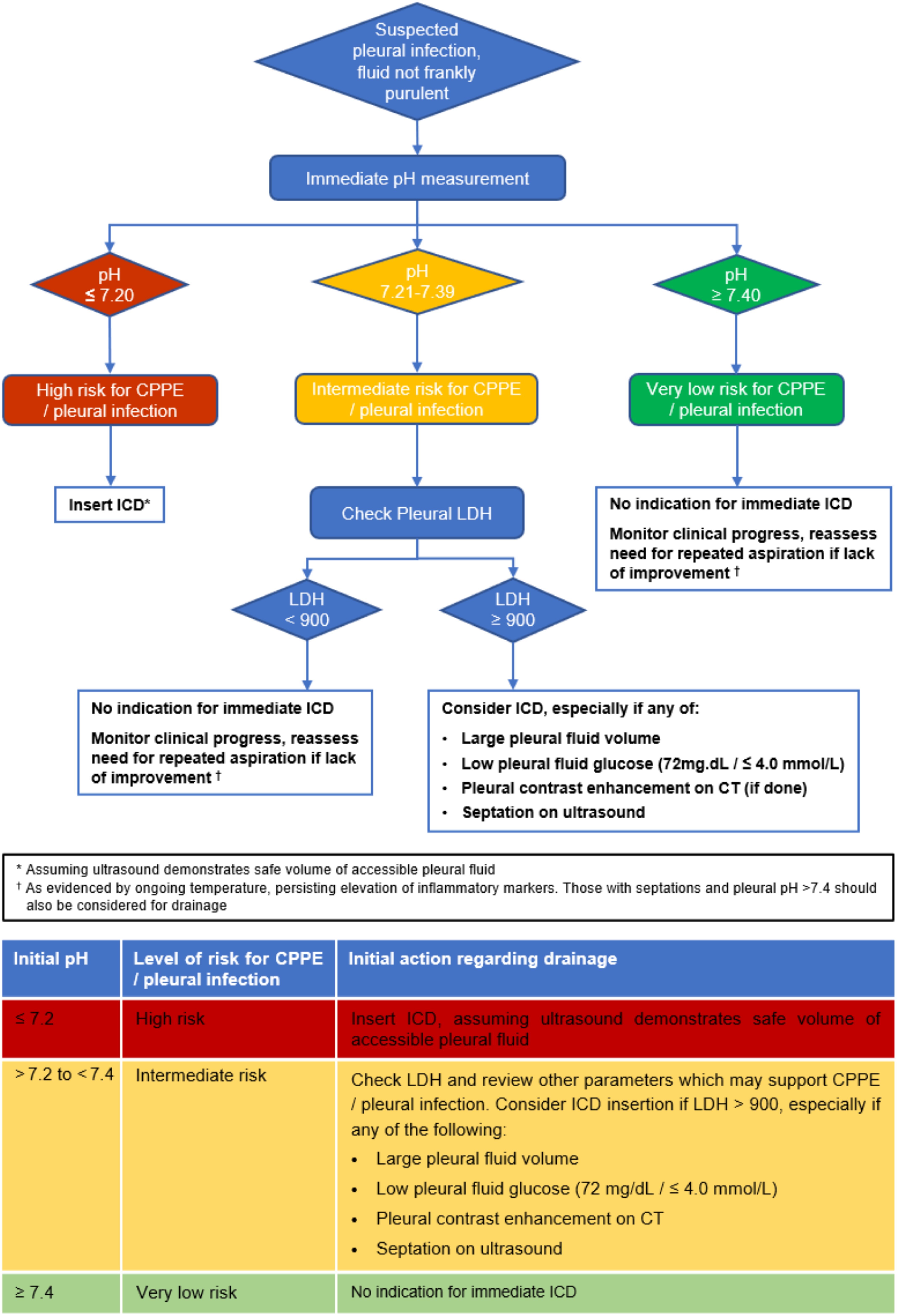
  • For patients with parapneumonic effusion (PPE) or suspected pleural infection, where diagnostic aspiration does not yield frank pus, immediate pH analysis should be performed. (Strong—by consensus)

  • For patients with suspected complex parapneumonic effusion (CPPE):

    • If pleural fluid pH is ≤7.2, this implies a high risk of CPPE or pleural infection and an intercostal drain (ICD) should be inserted if the volume of accessible pleural fluid on ultrasound makes it safe to do so. (Strong—by consensus)

    • If pleural fluid pH is >7.2 and <7.4, this implies an intermediate risk of CPPE or pleural infection. Pleural fluid lactate dehydrogenase should be measured and if >900 IU/L ICD should be considered, especially if other clinical parameters support CPPE (specifically ongoing temperature, high pleural fluid volume, low pleural fluid glucose (72 mg/dL ≤4.0 mmol/L), pleural contrast enhancement on CT or septation on ultrasound. (Strong—by consensus)

    • If pleural fluid pH is ≥7.4, this implies a low risk of CPPE or pleural infection and there is no indication for immediate drainage. (Strong—by consensus)

  • In the absence of readily available immediate pleural fluid pH measurement, an initial pleural fluid glucose <3.3 mmol/L may be used as an indicator of high probability of CPPE/pleural infection and can be used to inform decision to insert ICD in the appropriate clinical context. (Strong—by consensus)

Good practice points

  • Clinicians should be mindful of alternative diagnoses that can mimic PPE with a low pH and potential for loculations (eg, rheumatoid effusion, effusions due to advanced malignancy/mesothelioma).

  • Pleural fluid samples taken for pH measurement should not be contaminated with local anaesthetic or heparin (eg, by extruding all heparin from an arterial blood gas syringe) as this lowers pleural fluid pH. Delays in obtaining a pleural fluid pH or residual air in the sampling syringe will also increase pleural fluid pH.

  • In patients where a clinical decision is made not to insert an ICD at initial diagnostic aspiration, regular clinical reviews should be performed and repeat thoracocentesis considered to ensure that CPPE is not missed.

Optimal initial drainage strategy for established pleural infection

Recommendation

  • Initial drainage of pleural infection should be undertaken using a small bore chest tube (14 F or smaller). (Conditional—by consensus)

Good practice points

  • Due to the lack of supporting evidence, early surgical drainage under video-assisted thoracoscopy surgery (VATS) or thoracotomy should not be considered over chest tube (‘medical’) drainage for the initial treatment of pleural infection.

  • Due to lack of supporting evidence, medical thoracoscopy should not be considered as initial treatment for pleural infection.

Intrapleural therapy for managing pleural infection

Recommendations

  • Combination tissue plasminogen activator (TPA) and DNAse should be considered for the treatment of pleural infection, where initial chest tube drainage has ceased and leaves a residual pleural collection. (Conditional—by consensus)

  • Saline irrigation can be considered for the treatment of pleural infection when intrapleural TPA and DNase therapy or surgery is not suitable. (Conditional—by consensus)

  • Single agent TPA or DNAse should not be considered for treatment of pleural infection. (Conditional—by consensus)

  • Streptokinase should not be considered for treatment of pleural infection. (Conditional)

Good practice points

  • Patient consent should be taken when using TPA and DNase as there is a potential risk of bleeding.

  • When administering TPA plus DNase the regime should be 10 mg TPA twice daily (10 mg two times per day)+5 mg DNase two times per day for 3 days, based on randomised controlled trial data. Based on retrospective case series data, lower dose 5 mg TPA two times per day+5 mg DNase two times per day for 3 days may be as effective, and can be used if considered necessary.

  • Reduced doses of TPA may be considered in those with a potentially higher bleeding risk (eg, those on therapeutic anticoagulation which cannot be temporarily ceased).

  • For details on administration of intrapleural treatments, please refer to the British Thoracic Society (BTS) Clinical Statement on Pleural Procedures.(1)

Optimal surgical approach and surgical method for managing pleural infection

Recommendation

  • VATS access should be considered over thoracotomy for adults in the surgical management of pleural infection. (Conditional)

Good practice points

  • When selecting a surgical access for the treatment of pleural infection in adults, it is important to ensure the technique can facilitate optimal clearance of infected material and achieve lung re-expansion where appropriate.

  • Extent of surgery should be tailored according to patient and empyema stage when the lung is not completely trapped (drainage vs debridement).

  • Decortication should be a decision that is individualised to the patient with a trapped lung based on assessment of patient fitness and empyema stage.

iv - Pleural Malignancy

Optimal imaging modality for diagnosing pleural malignancy

Recommendations

  • Ultrasound may be a useful tool at presentation to support a diagnosis of pleural malignancy, particularly in the context of a pleural effusion, where appropriate sonographic skills are present. (Conditional)

  • CT allows assessment of the entire thorax, and positive findings may support a clinical diagnosis of pleural malignancy when biopsy is not an option (Conditional); however, a negative CT does not exclude malignancy. (Strong—by consensus)

  • PET-CT can be considered to support a diagnosis of pleural malignancy in adults when there are suspicious CT or clinical features and negative histological results, or when invasive sampling is not an option. (Conditional)

Good practice points

  • Imaging can play an important role in the assessment of pleural malignancy, but results should be interpreted in the context of clinical, histological and biochemical markers.

  • Features of malignancy may not be present on imaging at presentation. Unless a clear diagnosis is reached by other means (eg, biopsy), monitoring with follow-up imaging of patients presenting with pleural thickening and unexplained unilateral pleural effusion should be considered to exclude occult malignancy.

  • MRI has potential as a diagnostic tool in pleural malignancy. Its clinical value has yet to be determined and its use should be limited to highly selected cases and research studies at the present time.

Systemic therapy for reducing the need for definitive pleural intervention for malignant pleural effusion

Recommendation

  • Definitive pleural intervention should not be deferred until after systemic anticancer therapy (SACT). (Conditional—by consensus)

Managing malignant pleural effusion

Pleural aspiration with no pleurodesis agent versus talc slurry pleurodesis

Recommendation

  • Management of MPE using talc pleurodesis (or another method) is recommended in preference to repeated aspiration especially in those with a better prognosis, but the relative risks and benefits should be discussed with the patient. (Conditional—by consensus)

Good practice points

  • Decisions on the best treatment modality should be based on patient choice.

  • Informed decision-making should include the role of inpatient versus ambulatory management and the potential risk of requiring further pleural interventions.

Indwelling pleural catheter versus talc slurry pleurodesis

Recommendation

  • Patients without known non-expandable lung should be offered a choice of indwelling pleural catheter (IPC) or pleurodesis as first-line intervention in the management of MPE. The relative risks and benefits should be discussed with patients to individualise treatment choice. (Conditional)

Good practice points

  • The psychological implications and potential altered body image aspects of having a semi-permanent tube drain in situ should not be underestimated and must be considered prior to insertion.

  • All patients who have had an IPC inserted should be referred to the community nursing team on discharge for an early assessment of the wound site, symptom control, support with IPC drainage and removal of sutures.

  • Patients and their relatives should be supported to perform community drainage and complete a drainage diary if they feel able to do so, to promote independence and self-management.

  • Complications such as infection refractory to community management, suspected drain fracture, loculations or blockage with persistent breathlessness should be referred back to the primary pleural team for further assessment.

Thoracoscopy and talc poudrage pleurodesis versus chest drain and talc slurry pleurodesis

Recommendation

  • Talc slurry or talc poudrage may be offered to patients with MPE to control fluid and reduce the need for repeated procedures. (Conditional)

Good practice points

  • Where a diagnostic procedure is being conducted at thoracoscopy (pleural biopsies), if talc pleurodesis is reasonable, this should be conducted during the same procedure via poudrage.

Surgical pleurodesis, or surgical decortication versus talc slurry pleurodesis

  • In selected patients considered fit enough for surgery, either surgical talc pleurodesis or medical talc slurry can be considered for the management of patients with MPE. The relative risks, benefits and availability of both techniques should be discussed with patients to individualise treatment choice. (Conditional—by consensus)

Good practice points

  • Informed decision-making should include the role of surgery versus ambulatory management with an IPC for the management of MPE in selected patients.

  • Decortication surgery may improve pleurodesis success in patients wih MPE with non-expandable lung, but the risks and benefits of IPC and surgical treatment should be discussed with patients, and treatment individualised according to circumstances (eg, fitness to undergo thoracic surgery).

Managing malignant pleural effusion and non-expandable lung

Pleural aspiration, talc slurry pleurodesis, talc poudrage pleurodesis, decortication surgery or indwelling pleural catheter

Good practice points

  • Decisions on treatment modality for MPE and non-expanded lung should be based on patient choice, with the relative risks and benefits of each modality discussed with the patient, but patients should be made aware of the limited evidence base regarding treatment options for non-expandable lung.

  • IPCs are effective at controlling symptoms in non-expandable lung and should be considered, but it may be appropriate to undertake pleural aspiration first to assess symptomatic response.

  • Pleural aspiration may result in a need for multiple procedures so alternatives should be discussed with the patient.

  • In patients with radiologically significant (>25%) non-expandable lung requiring intervention for a symptomatic MPE, current evidence suggests the use of an IPC rather than talc pleurodesis.

  • In patients with MPE and <25% non-expandable lung, talc slurry pleurodesis may improve quality of life, chest pain, breathlessness and pleurodesis rates.

  • Decortication surgery may improve pleurodesis success in selected patients with MPE and non-expandable lung, but the risks and benefits of IPC and surgical treatment should be discussed with patients, and treatment individualised according to circumstances (eg, fitness to undergo thoracic surgery).

Managing malignant pleural effusion and septated effusion (on radiology)

Intrapleural enzymes versus surgery, or no treatment

Good practice points

  • Intrapleural fibrinolytics can be considered in highly selected symptomatic patients with MPE and septated effusion to try to improve breathlessness.

  • Intrapleural fibrinolytics may be used in patients with MPE and septated effusion and an IPC to improve drainage if flushing the IPC with normal saline or heparin saline does not improve drainage.

  • Surgery can be considered for palliation of symptoms in a minority of patients with significantly septated MPE and associated symptoms and otherwise good prognosis and performance status.

Managing malignant pleural effusion treated with an indwelling pleural catheter

Symptom-based/conservative drainage versus daily drainage

Recommendations

  • Where IPC removal is a priority, daily IPC drainages are recommended to offer increased rates of pleurodesis when compared with less frequent drainages of symptom-guided or alternate drainage regimes. (Conditional)

  • Patients should be advised that they do not require daily drainage to control symptoms of breathlessness and chest pain if they wish to opt for a less intensive regime. (Strong—by consensus)

Good practice points

  • Decisions on the optimal drainage frequency should be based on patient choice.

  • Informed decision-making should include the explanation of the effect of drainage regimes on the patient-centre outcomes such as breathlessness and the possibility of autopleurodesis during the disease course.

  • Although daily drainage may result in earlier removal of IPC, there may be an associated cost associated with the increased number of drainage events (both to the healthcare system and to the patient). This has been addressed in a modelling study (2) and should be considered.

Intrapleural agents (talc or other pleurodesis agents)

Recommendation

  • Instillation of talc via an IPC should be offered to patients with expandable lung where the clinician or patient deems achieving pleurodesis and IPC removal to be important. (Conditional—by consensus)

Intrapleural chemotherapy versus systemic treatment for treating pleural malignancy

Recommendation

  • Intrapleural chemotherapy should not be routinely used for the treatment of MPE. (Conditional—by consensus)

Good practice point

  • All patients of good performance status with metastatic malignancy should be considered for SACT as standard of care as per national guidelines.

Using prognostic or predictive scores to provide prognostic information for patients with malignant pleural effusion

Good practice points

  • Clinicians may consider using a validated risk score for MPE, if the information is of use in planning treatments or in discussion with patients.

  • Patients with pleural malignancy should be managed in a multidisciplinary way, including referral to specialist palliative care services where appropriate.

Clinical pathways

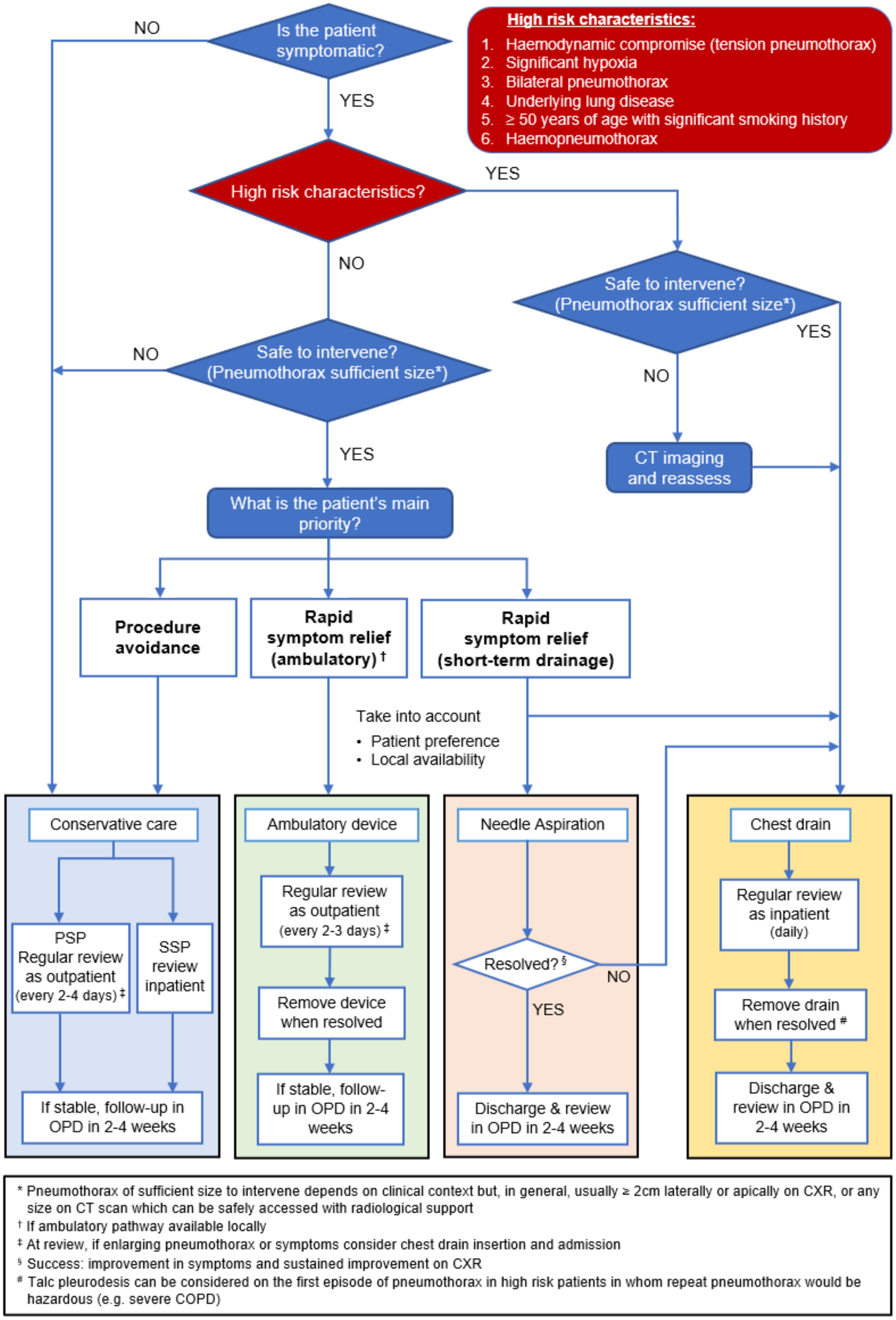
Pneumothorax pathway

Unilateral pleural effusion disagnostic pathway


Table 1

Light’s criteria

Pleural fluid is an exudate if one or more of the following criteria are met: 

•      Pleural fluid protein divided by serum protein is >0.5

•      Pleural fluid lactate dehydrogenase (LDH) divided by serum LDH is >0.6

•      Pleural fluid LDH >2/3 the upper limits of laboratory normal value for serum LDH


Table 2

Transudates

Exudates

Common

Common

•      Congestive cardiac failure

•      Malignancy

•      Liver cirrhosis

•      Pleural infection

•      Hypoalbuminaemia

•      Pulmonary embolism

•      Nephrotic syndrome

•      Autoimmune pleuritis

Less common

Less common

•      Nephrotic syndrome

•      Drugs

•      Mitral stenosis

•      Lymphatic disorders

•      Peritoneal dialysis

•      Meigs syndrome

•      Chronic hypothyroidism

•      Post-coronary artery bypass graft

•      Constrictive pericarditis

•      Benign asbestos related pleural effusion


Table 3

Causes of lymphocytic pleural effusion

Malignancy

Tuberculosis

Lymphoma

Congestive cardiac failure

Post-coronary bypass graft

Rheumatoid arthritis

Chylothorax

Yellow nail syndrome


Table 4

Causes of bilateral pleural effusions

Congestive cardiac failure

Hypoalbuminaemia

Renal failure

Liver failure

SLE and other autoimmune diseases

Widespread malignancy including abdominal/pelvic malignancy

Bilateral pulmonary embolus


Table 5

Pleural fluid lipid values in chylothorax and pseudochylothorax


Chylothorax:


Triglycerides

high >1.24 mmol/L (110 mg/dL)

Cholesterol

low

Cholesterol crystals

absent

Chylomicrons

usually present

Pseudochylothorax:


Triglycerides

low

Cholesterol

high >5.18 mmol/L (200 mg/dL)

Cholesterol crystals

often present

Chylomicrons

absent


Table 6


Chylothorax


•      Trauma:

thoracic surgery (especially if involving posterior mediastinum, eg, oesophagectomy), thoracic injuries

•      Neoplasm:

lymphoma or metastatic carcinoma

•      Miscellaneous:

disorders of lymphatics (including lymphangioleiomyomatosis), tuberculosis, cirrhosis, obstruction of the central veins, chyloascites

•       Idiopathic (about 10%)

Pseudochylothorax:

•      Tuberculosis

•      Rheumatoid arthritis

Suspected pleural infection, non-purulent fluid - initial decision tree

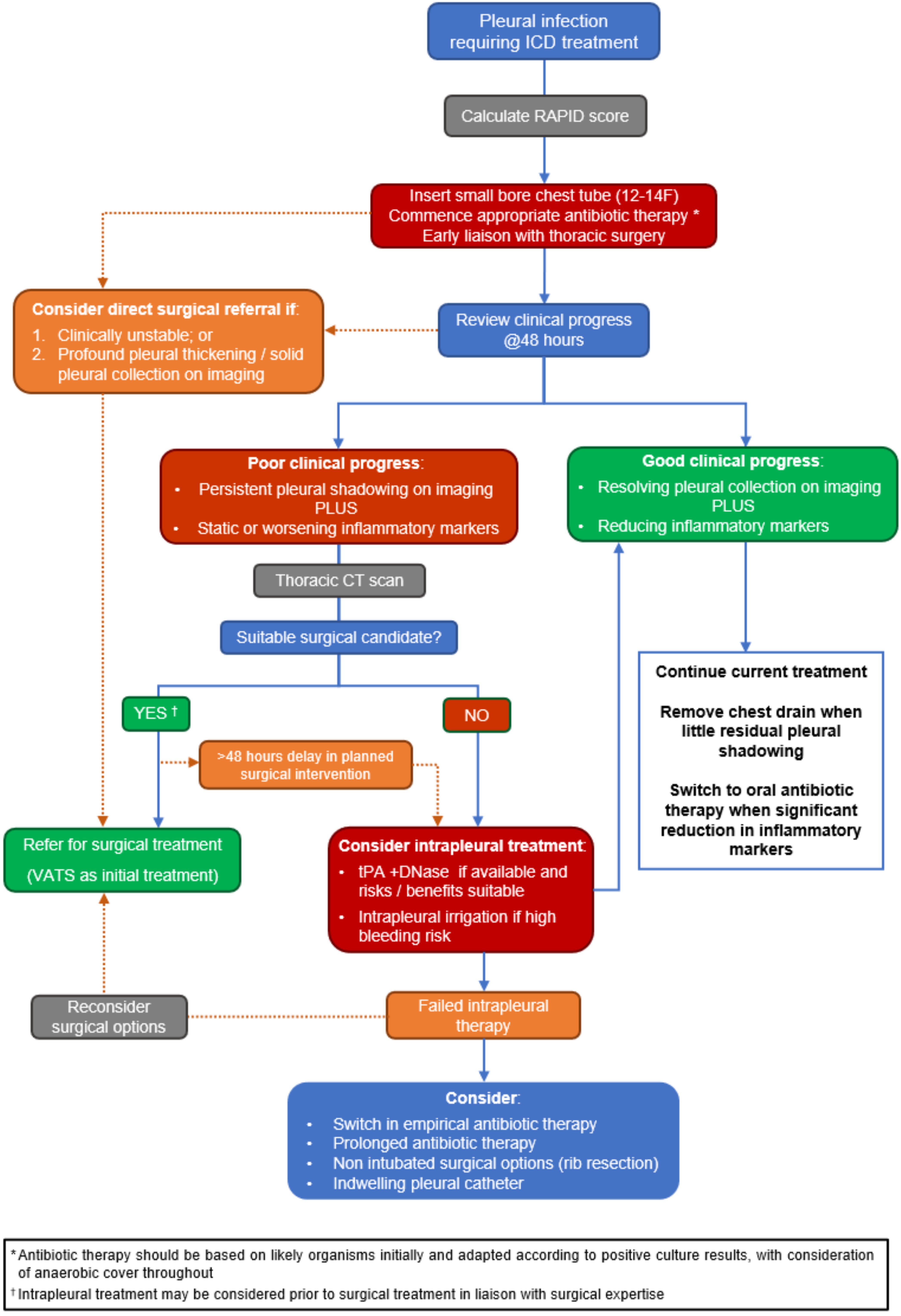
Pleural infection treatment pathway

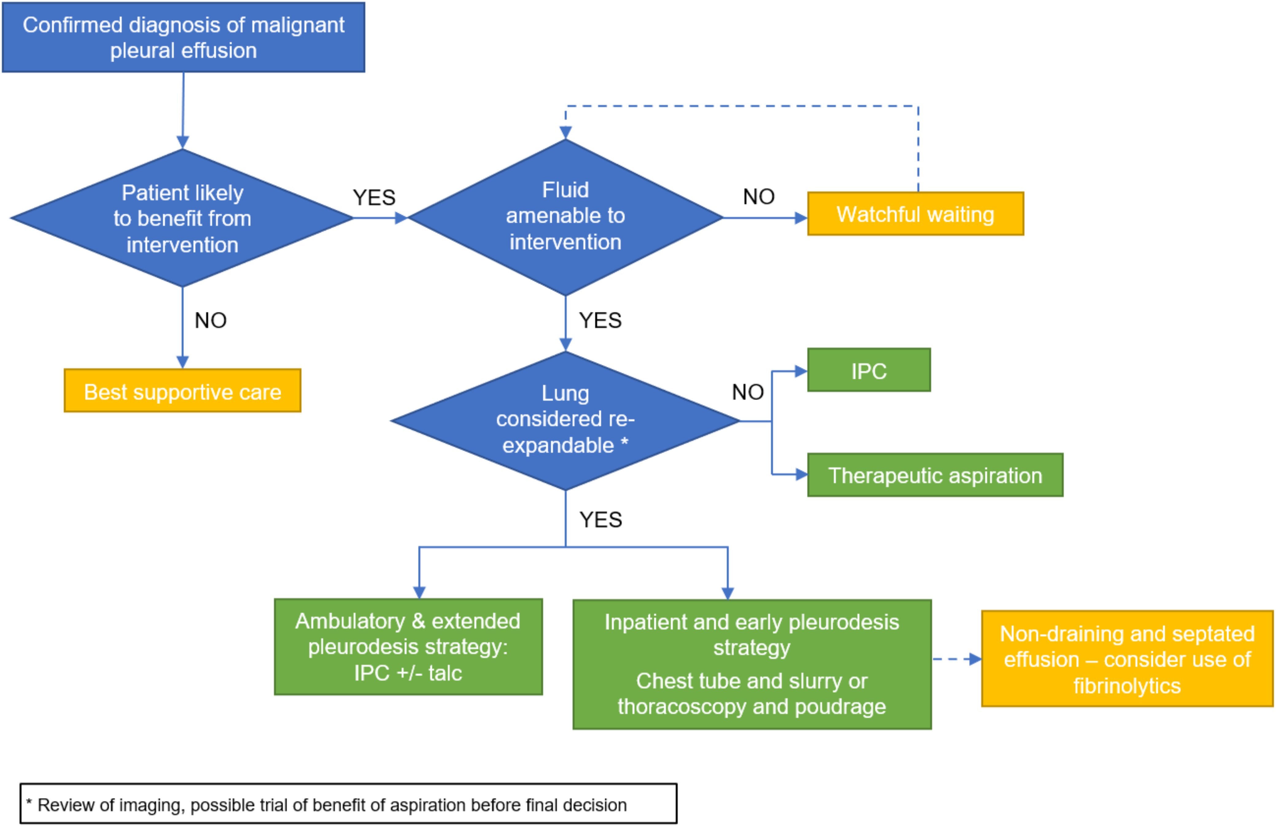
Malignant pleural effusion pathway

Glossary

Glossary and acronyms

AAFB Acid alcohol fast bacilli 
ADA Adenosine deaminase
ANA Antinuclear antibody
bd bis die (to be taken twice times daily)
CA15-3  Cancer antigen 15-3
CA19-9     Carbohydrate antigen 19-9
CA72-4     Cancer antigen 72-4
CEA  Carcinoembryonic antigen
CDSR Cochrane database of systematic reviews
CENTRAL  Cochrane central register of controlled trials 
CI Confidence intervals
COPD  Chronic obstructive pulmonary disease
CPPE Complicated/complex parapneumonic effusion
CRP C-reactive protein
CT Computed tomography
CXR Chest x-ray
CYFRA 21-1 Fragment of cytokeratin 19
DVT Deep vein thrombosis
GDG Guideline development group
GPP Good practice point 
GRADE  Grading of recommendations, assessment, development and evaluation
ICD Intercostal chest drain/drainage 
IFN-gamma Interferon gamma
IPC Indwelling pleural catheter
IgG4 Immunogobulin G4
LDH Lactate dehydrogenase 
LOS Length of hospital stay
LVEF Left ventricular ejection fraction
MPE Malignant pleural effusion
MPM Malignant pleural mesothelioma
MRI Magnetic resonance imaging
MRSA Methicillin-resistant Staphylococcus aureus
NA Needle aspiration 
NEL Non-expandable lung 
NSCLC Non-small-cell lung carcinoma 
NT-proBNP N-terminal pro hormone pro-brain natriuretic peptide
PET-CT Positron emission tomography-computed tomography 
PICO Population, intervention, comparator and outcome
PPE Parapneumonic effusions (PPE) 
PSP Primary spontaneous pneumothorax 
QoL Quality of life
RPO Re-expansion pulmonary oedema 
SACT Systemic anti-cancer therapy
SP  Spontaneous pneumothorax
SSP Secondary spontaneous pneumothorax
TB Tuberculosis
TDS Ter die sumendum (to be taken three times daily)
TPA Tissue plasminogen activator
TPE Tuberculous pleural effusion
TUS Thoracic ultrasound 
UPPE Uncomplicated parapneumonic effusion
VATS Video-assisted thoracoscopy surgery 
VEGF Vascular endothelial growth factor


Scope

Aim of the guideline

This guideline aims to provide evidence-based guidance on the
investigation and management of:

i. Spontaneous pneumothorax (SP)
ii. Undiagnosed unilateral pleural effusion
iii. Pleural infection
iv. Pleural malignancy

Pleural disease is common and represents a major and rapidly
developing subspecialty that presents to many different hospital
services. Since the last British Thoracic Society (BTS) Guideline for pleural disease published in 2010,3–9 many high-quality and practice changing studies, using patient-centred outcomes,  have been published. The paradigms for the investigation and management of pleural disease have therefore shifted. For example, ambulatory treatments have become much more prominent in the management of pleural disease. 

This guideline aims to capture this evidence and use it to answer the most important questions relevant to today’s practice.

Intended users of the guideline and target populations

The guideline will be of interest to UK-based clinicians caring for adults with pleural disease, including chest physicians, respiratory trainees, specialist respiratory nurses, specialist lung cancer nurses, specialist pleural disease nurses, pathologists, thoracic surgeons, thoracic surgeon trainees, acute physicians, oncologists, emergency physicians, hospital practitioners, intensive care physicians, palliative care physicians, radiologists, other allied health professional and patients and carers.

Guideline group members were selected to offer a broad geographical coverage of the UK and to include specialists with backgrounds in respiratory medicine, thoracic surgery, oncology, palliative care, nursing and pathology. The group included specialists from tertiary centres as well as district general hospitals.

Scope of the guideline

The guideline is specifically designed to answer important questions in the investigation and management of pleural disease in adults.

Questions have been agreed by the whole guideline group. While as many important questions as possible have been included, there are areas that have not been covered. As this guideline covers four broad areas of pleural disease, the number of questions is limited by the practicalities of writing a guideline with a large scope that remains relevant and up to date at the point of publication and a workload manageable by the guideline group.

This guideline covers adult patients in both inpatient and ambulatory settings, and questions from investigation to management in the inpatient and outpatient settings and by specialists of all disciplines involved in the care of patients with pleural disease.

Areas not covered by the Guideline

Mesothelioma has been excluded from this guideline as this is already covered in the BTS Guideline for the investigation and management of pleural mesothelioma. (10) Benign (non-infectious, non-pneumothorax) pleural disease and rare pleural diseases are also excluded. Guidance on pleural interventions are covered in the BTS Clinical Statement on Pleural Procedures.(1)

Limitations of the guideline

Healthcare providers need to use clinical judgement, knowledge and expertise when deciding whether it is appropriate to apply recommendations for the management of patients. The recommendations cited here are a guide and may not be appropriate for use in all situations. The guidance provided does not override the responsibility of healthcare professionals to make decisions appropriate to the circumstances of each patient, in consultation with the patient and/or their guardian or carer.

Members of the Guideline Development Group

The Guideline Development Group (GDG) was chaired by three respiratory consultants—Professor Nick Maskell, Professor Najib Rahman and Dr Mark Roberts. The GDG had a wide membership and included colleagues from respiratory medicine, oncology, radiology, pathology and palliative medicine. Two patient representatives were recruited to the group, but due to personal circumstances both had to withdraw before completion of the guideline (August 2019 and July 2021). However, two further patient representatives were recruited at the end of the guideline process to review the final guideline and provide the patients’ perspective.

Those on the group were not required to be BTS members and a full list of members can be seen in Appendix 2

Guideline questions

The guideline is specifically designed to answer important questions in the investigation and management of pleural disease in adults. Questions have been agreed by the whole guideline group. While as many important questions as possible have been included, there are areas that have not been covered. As this guideline covers four broad areas of pleural disease, the number of questions is limited by the practicalities of writing a guideline with a large scope that remains relevant and up to date at the point of publication and a workload manageable by the guideline group.

This guideline covers adult patients in both inpatient and ambulatory settings, and questions from investigation to management in the inpatient and outpatient settings and by specialists of all disciplines involved in the care of patients with pleural disease.

Methodology

Establishment of GDG

The GDG was convened in July 2018, with the first meeting taking place in November 2018. The full GDG met 10 times during the development of the guideline and kept in close contact by teleconference and email throughout the process.

Methodology of guideline production

This BTS Guideline uses Grading of Recommendations, Assessment, Development and Evaluation (GRADE) methodology in the guideline development process. Full details are provided in the BTS Guideline production manual.

Summary of key questions, outcomes and literature search

Clinical questions were defined from the scope of the guideline and formulated into systematic review type questions (diagnostic accuracy, intervention or prognostic) according to the nature of the question. A full list of clinical questions for each section of the guideline is provided below. 

Patient-centred outcomes were agreed by the group for each question.

The Population, Intervention, Comparator and Outcome (PICO) framework, or equivalent for the diagnostic accuracy and prognostic review questions, formed the basis of the literature search. The initial searches were completed by the University of York (and latterly by BTS Head Office). Systematic electronic database searches were conducted to identify all papers that may be relevant to the guideline. For each question, the following databases were searched: Cochrane Database of Systematic Reviews, Cochrane Central Register of Controlled Trials, MEDLINE and EMBASE. The search strategy is available for review below.

Literature review

Two literature searches were conducted for the guideline, with the number of resulting abstracts from each search shown in Table 1.

Table 1: Literature searches were conducted for each section of the guideline as follows:

Section

Search 1 date

Number of abstracts

Search 2 date

Number of abstracts

Spontaneous pneumothorax

20 Mar 2020

  6325

18 May 2021

1260

Investigation of the undiagnosed unilateral pleural effusion

18 Mar 2019

  6773

13 May 2021

2199

Pleural infection

17 Dec 2019

  4138

20 May 2021

 822

Pleural malignancy 

03 Apr 2019

14276

11 May 2021

3641


Letters, conference papers and news articles were removed and criteria for initial screening of the abstracts were:

  • Does the study type match the study type criteria in the clinical question protocols?

  • Does the population match the clinical question population(s)?

  • Is the abstract in English?

The remaining abstracts were screened by Professor Maskell, Professor Rahman and Dr Roberts and potentially relevant abstracts allocated to the relevant clinical questions. Abstracts were not rejected on the basis of the journal of publication, authorship or country of origin.

GDG members were allocated to work on individual questions in small groups. Each abstract was read and at least two members agreed whether the abstract was ‘potentially relevant’ or ‘not relevant’ to the clinical question of interest. Abstracts were excluded if they were deemed ‘not relevant’ to the clinical question.

Full papers were obtained for all abstracts assigned as ‘potentially relevant’. Each full paper was reviewed to assess if it addressed:

  1. The clinical question population;

  2. The index test and reference standard (for diagnostic accuracy questions), the intervention and comparator (for intervention questions) or the exposure and referent (for prognostic questions);

  3. The study type(s) defined in the clinical question protocol;

  4. The clinical question outcome(s).

Each full paper fulfilling the above criteria, and agreed by at least two members of the GDG, was ‘accepted’ for meta-analysis and subsequent critical appraisal.

In circumstances where there was little, or no supporting evidence that fulfilled the above criteria, the full paper inclusion strategy was widened to include evidence that partially addressed the clinical question.

The second literature search (Search 2, Table 1) was undertaken in May 2021 to capture additional published evidence while the guideline was in development prior to finalising the draft document. The additional abstracts were reviewed and allocated to the clinical questions as above.

The full list of abstracts has been retained and is kept in an archive.

Systematic review of evidence

Each ‘accepted’ full paper underwent a systematic review. Data were extracted and meta-analyses were performed for each clinical question on an outcome-by-outcome basis for intervention reviews, or an index test basis for diagnostic accuracy reviews. If meta-analysis was not possible, for example, if there was insufficient evidence to perform a meta-analysis, if data could not be extracted to input into a meta-analysis, or data across studies had been published in different formats, all relevant supporting data were tabulated where possible.

All full papers contributing towards a meta-analysis underwent critical appraisal. For all non-meta-analysed data included in an evidence review, contributing papers also underwent critical appraisal where possible.

Meta-analyses and risk of bias assessments (critical appraisal) were performed in Review Manager V.5.3 and agreed by at least two members of the GDG. Diagnostic accuracy meta-analyses involved an additional step which was performed by BTS Head Office using the MetaDTA app.(11) Papers no longer deemed relevant were removed from the systematic review, with the decision to ‘exclude’ a paper solely based on it not fulfilling the clinical question criteria.

GRADE analysis of the evidence

Having generated evidence profiles for each of the clinical question, GDG question groups assessed the quality of the evidence using the GRADE methodology.(12) Where meta-analysis was not possible, but studies had used comparable methodologies and data reporting methods to allow an assessment of the quality of the data, a prognostic review GRADE analysis approach was used.(13-14)

Where GRADE analysis was not possible, but GDG members felt the evidence was important to be included in the evidence statements, these have been listed as (Ungraded). Definitions of the evidence statement (GRADE) scores are shown in Table 2.

Table 2: Evidence statement (GRADE) score definitions

GRADE

Definition

High

High confidence that the true effect is close to the estimated effect

Moderate

Logo

Description automatically generated

Moderate confidence that the true effect is close to the estimated effect

Low

Shape

Description automatically generated with medium confidence

Low confidence that the true effect is close to the estimated effect

Very Low

Very low confidence that the true effect is close to the estimated effect

Ungraded


Where GRADE analysis was not possible, due to differences in data format across the presented evidence, but the evidence was deemed important by the GDG


Each clinical question review was reviewed by the full GDG during the regular meetings and consensus was reached in relation to the evidence summary.

Recommendation development

The GDG proceeded to decide on the direction and strength of recommendations considering the quality of the evidence, the balance of desirable and undesirable outcomes and the values and preferences of patients and others. GRADE specifies two categories of strength for a recommendation, as shown in Table 3.

Table 3: Explanation of the terminology used in BTS recommendations

Strength

Benefits and risks

Implications

Strong 

Recommended, so “offer”

Benefits appear to outweigh the risks (or vice versa) for the majority of the target group

Most service users would want to, or should receive this intervention

Conditional

Suggested, so “consider”

Risks and benefits are more closely balanced, or there is more uncertainty in likely service users’ values and preferences

Service users should be supported to arrive at a decision based on their values and preferences


From the outset, it was acknowledged that there would be little high-quality evidence for some of the clinical questions identified. In this instance, low-grade evidence was considered, along with the expert opinion of the GDG via informal consensus at the meetings.

Good practice points (GPPs) were developed by informal consensus in areas where there was no quality evidence, but the GDG felt that some guidance, based on the clinical experience of the GDG, might be helpful to the reader. These are indicated as shown below.

  • Advised best practice based on the clinical experience of the GDG.

In some instances where evidence was limited, but GDG members felt that it was important to include a recommendation rather than a GPP, recommendations were agreed by informal consensus and categorised as (Strong—by consensus) or (Conditional—by consensus), based on the same criteria detailed in Table 3.

Cost-effectiveness was not considered in detail as in-depth economic analysis of recommendations falls outside of the scope of the BTS Guideline production process. However, the GDG were asked to be mindful of any potential economic barriers to the implementation of recommendations and GPPs.

Research recommendations were also identified and are detailed in online supplemental appendix 2.

Drafting/reviewing the guideline

The guideline group corresponded regularly by email and meetings of the full group were also held in the period between November 2018 and late 2020. A revised draft guideline document was circulated to all the relevant stakeholders for consultation in May 2022 followed by a period of online consultation.

The BTS Standards of Care Committee reviewed the ‘Investigation of the undiagnosed unilateral pleural effusion’ and ‘Pleural malignancy’ sections of the draft guideline in March 2020 and the full guideline in March 2022.

Main guideline

The Guideline and Summary can also be accessed via the Thorax journal here: Full Guideline and Summary.

Spontaneous pneumothorax

Introduction

The term pneumothorax describes air in the pleural space and is characterised as spontaneous in the absence of trauma or causative medical intervention. It is an increasing problem, with annual hospital admission rates rising from 9.1 to 14.1 per 100 000 population in the last 50 years, leading to substantial symptom burden and healthcare utilisation.(15 16) Since the last version of the BTS pneumothorax guideline, published in 2010 (5) there have been several large high-quality clinical trials examining the management of SP.(17–20)

This guideline seeks to consolidate and update the pneumothorax guidelines in the light of this subsequent research using GRADE methodology and addresses the following clinical questions addressing adults with pneumothorax:

  • What is the best acute management for SP? (Question A1)

  • What is the optimal management of patients after resolution of first episode of pneumothorax? (Question A2)

  • What is the optimal management of patients with ongoing air leak? (Question A3)

  • What is the optimal surgical approach when performing surgery? (Question A4)

  • What is the optimal operation when performing surgery? (Question A5)

Other areas of clinical importance that are not covered by the guideline questions are discussed in the ‘Other areas of clinical importance not covered by the clinical questions’ section, including traumatic and iatrogenic pneumothorax which are not specifically covered in the evidence review.

Definitions and treatment principles

Spontaneous pneumothoraces can be subclassified into primary spontaneous pneumothorax (PSP) in the absence of suspected lung disease or secondary spontaneous pneumothorax (SSP) in patients with established underlying lung disease. This distinction does not imply that patients with PSP have normal underlying lung parenchyma, with the majority demonstrating emphysema-like pulmonary changes on CT imaging, but instead reflects that current management and outcomes differ between the two patient groups. Patients can also be characterised as SSP if they are older than 50 years of age and have a smoking history. This categorisation reflects case series data that this cohort may respond differently to needle aspiration (NA) than younger patients or non-smokers.

There have been substantial changes in recommendations in this BTS Guideline compared with the 2010 Guidelines. Size of pneumothorax is no longer an indication for invasive management (although does dictate the safety of conducting an intervention) and the use of chest drains is mainly centred around patients with high-risk characteristics (Appendix 1, Pneumothorax pathway). The expanded evidence base now allows for a more personalised approach and greater patient choice. For details of interventions and how these are best conducted, please refer to the BTS Clinical Statement on Pleural Procedures for further details.(1)

What is the best acute management for spontaneous pneumothorax?

Drainage of symptomatic pneumothorax, either with NA or intercostal chest drain (ICD) attached to an underwater seal is the current standard of care for PSP. There is ongoing debate over the respective benefits of NA over ICD, with multiple recent randomised trials comparing NA with ICD. Conservative management (ie, no active intervention) is often undertaken in patients with small or incidental PSP, but could be an alternative to NA or chest drain in patients with larger pneumothoraces.

Ambulatory treatment using a purpose-made device containing a one-way valve, or Heimlich valve attached to chest drain has the potential to allow outpatient management of pneumothorax. A proportion of pneumothoraces will recur and both chemical pleurodesis via chest tube and thoracic surgery have the potential to reduce this risk. Thoracic surgery is often the treatment of choice for ongoing air leak, or for those with recurrent pneumothorax. However due to the risk of recurrence, trials have been performed to establish whether thoracic surgery could be offered at first presentation of pneumothorax. Conservative management, in which no intervention is undertaken and the patient is observed or reviewed repeatedly, is also a further alternate initial potential treatment strategy. Hence, the first clinical question was:

A1 For adults with spontaneous pneumothorax, is conservative management, needle aspiration, ambulatory management, chemical pleurodesis or thoracic surgery better than intercostal drainage at improving clinical outcomes?

A summary of the evidence review is shown in table 4 and the evidence statements (conclusions from the evidence review), recommendations and GPPs are presented below.

The full evidence review is available in online supplemental appendix A1.

Table 4 Evidence Review Summary for 'What is the best acute management for spontaneous pneumothorax?

Clinical outcomes

Summary of Evidence review (treatment versus ICD) [95% CI]

(Treatment)

Conservative management

Needle aspiration (NA)

Ambulatory management

Chemical pleurodesis

Thoracic surgery

Length of hospital stay (LOS)


Shortened LOS with conservative management *

2.55 days shorter [2.24 to 2.87] with NA (PSP) †

3.47 days shorter [2.20 to 4.73] with ambulatory management †

No difference

No difference

Pneumothorax recurrence

Lesser risk with conservative management (111/1000 [80 to 155]) compared with (179/1000) †

No difference

No difference

Lesser risk with chemical pleurodesis (179/1000 [138 to 227]) compared with 320/100 (PSP and SSP) †

Lesser risk with thoracic surgery (54/1000 [36 to 80]) compared with 298/1000 (PSP and SSP) †

Re-admission

Not enough evidence

Not reported

No difference

Not reported

Not reported

Need for further pleural procedures

Not enough evidence

Greater need with NA (626/1000 [544 to 719] compared with 240/1000

No difference

Not reported

Not reported

Complications

Reduced post-treatment complications with conservative management *

No overall difference in complications, but may be an reduction in subcutaneous emphysema following NA (9/1000 [1 to 70]) compared with 92/1000

No difference

Not enough evidence

No difference

Pain and breathlessness

Not enough evidence

Not enough evidence

Not enough evidence
 

Greater need for opioids with chemical pleurodesis *

Not reported

Quality of life (QoL)

Not enough evidence

Not reported

Not reported

Not reported

Not reported

Mortality

Not reported

Not reported

Not reported

Not reported

No difference

*Meta-analysis not possible, data reported in different formats.

†Meta-analysis results reported as per 1000 patients.

ICD, intercostal drainage; LOS, length of stay; NA, needle aspiration; PSP, primary spontaneous pneumothorax; SSP, secondary spontaneous pneumothorax.

Evidence statements

Conservative Management
  • Length of hospital stay appears to be shorter following conservative management for the treatment of PSP in adults when compared with ICD. (Ungraded)

  • Risk of pneumothorax recurrence appears to be greater following ICD when compared with conservative management for the treatment of PSP in adults. (Very low)

  • There may be more complications experienced following ICD when compared with conservative management for the treatment of PSP in adults. (Ungraded)

Needle aspiration
  • Length of hospital stay appears to be shorter following NA for the treatment of PSP in adults when compared with ICD. (Low)

  • There appears to be no difference in the rate of recurrence between NA or ICD for the treatment of PSP in adults. (Very low)

  • There appears to be a greater need for further pleural procedures following NA when compared with ICD for the treatment of PSP in adults. (Very low)

  • The risk of overall complications following NA or ICD appear to be the same for the treatment of PSP in adults (Very low), but there may an increased risk of subcutaneous emphysema following ICD. (Low)

Ambulatory management
  • There appears to be a reduction in the length of hospital stay following ambulatory management when compared with standard care for the treatment of PSP in adults. (Moderate)

  • There appears to be no difference in the rate of pneumothorax recurrence, the rate of hospital re-admission, the need for pleural procedures or complications following ambulatory management or standard care for the treatment of PSP in adults. (Very low)

Chemical pleurodesis
  • There appears to be no difference in the length of hospital stay following chemical pleurodesis or ICD alone for the treatment of PSP in adults. (Low)

  • The risk of pneumothorax recurrence appears to be lower following chemical pleurodesis when compared with ICD alone for the treatment of PSP or SSP in adults. (Very low)

  • There appears to be a greater need for opioid pain relief following chemical pleurodesis when compared with ICD alone for the treatment of PSP in adults. (Moderate)

  • Although there appears to be no difference in mortality rate at time of treatment (Very low), tetracycline chemical pleurodesis may cause greater post-treatment mortality when compared with ICD alone for the treatment of pneumothorax in adults. (Very low)

Thoracic surgery
  • Length of hospital stay appears to be shorter following thoracic surgery, when compared with ICD, for the treatment of PSP in adults. (Very low)

  • The rate of pneumothorax recurrence appears to be reduced following thoracic surgery, when compared with ICD, for the treatment of PSP in adults. (Very low)

  • Pneumonia and persistent air leak complications appear to be greater following video-assisted thoracic surgery (VATS), when compared with ICD, for the treatment of PSP in adults. (Very low)

  • There appears to be no difference in the rate of mortality following thoracic surgery or ICD, for the treatment of pneumothorax in adults, with the mortality rate being very low for both treatments. (Very low)

Recommendations

  • Conservative management can be considered for the treatment of minimally symptomatic (ie, no significant pain or breathlessness and no physiological compromise) or asymptomatic PSP in adults regardless of size. (Conditional—by consensus)

  • Ambulatory management should be considered for the initial treatment of PSP in adults with good support, and in centres with available expertise and follow-up facilities. (Conditional)

  • In patients not deemed suitable for conservative or ambulatory management, NA or tube drainage should be considered for the initial treatment of PSP in adults. (Conditional)

  • Chemical pleurodesis can be considered for the prevention of recurrent SSP in adults (eg, patients with severe chronic obstructive pulmonary disease who significantly decompensated in the presence of a pneumothorax, even during/after the first episode). (Conditional)

  • Thoracic surgery can be considered for the treatment of pneumothorax in adults at initial presentation if recurrence prevention is deemed important (eg, patients presenting with tension pneumothorax, or those in high-risk occupations). (Conditional)

Good practice points

  • When establishing local ambulatory treatment pathways, planning and coordination between with the emergency department, general medicine and respiratory medicine is vital.

  • When performing chemical pleurodesis for the treatment of pneumothorax in adults, adequate analgesia should be provided before and after treatment.

  • All treatment options should be discussed with the patient to determine their main priority, with consideration for the least invasive option.

What is the optimal management of patients after resolution of a first episode of pneumothorax?

Recurrence following SP is a frequent concern and overall occurs in 32% of patients after a single episode of PSP(21) and 13%–39% after a first episode of SSP.(16) Current usual practice in the UK is to consider surgical intervention after the second episode of an SP to reduce subsequent further recurrences. The aim of the next question was to assess if there was evidence to support the use of surgical intervention (surgical pleurodesis or bullectomy) at an earlier stage in an elective context, prior to the first recurrence, comparing against non-surgical techniques (non-surgical talc or conservative management):

A2 What is the optimal management of patients after resolution of a first episode of pneumothorax?

The evidence statement and GPPs are presented below; and the full evidence review is presented in online supplemental appendix A2.

Evidence statement

There was no evidence relevant to the review.

Recommendations

Due to the lack of supporting evidence, no recommendations can be made on the role of elective surgery at an earlier stage to prevent recurrence.

Good practice points

  • Elective surgery may be considered for patients in whom recurrence prevention is deemed important (eg, at-risk professionals (divers, airline pilots, military personnel), or those who developed a tension pneumothorax at first episode).

  • Elective surgery should be considered for patients with a second ipsilateral or first contralateral pneumothorax.

  • Discharge and activity advice should be given to all patients post pneumothorax.

Discharge advice, flying and activity

All patients discharged after active treatment or otherwise should be given verbal and written advice to return to the accident and emergency department immediately should they develop further breathlessness. It is recommended that all patients should be followed up by a respiratory physician to ensure resolution of the pneumothorax, to institute optimal care of any underlying lung disease, to explain the risk of recurrence and the possible later need for surgical intervention and to reinforce lifestyle advice on issues such as smoking and air travel. Those managed by observation alone or by NA should be advised to return for a follow-up chest X-ray (CXR) after 2–4 weeks to monitor resolution. Patients managed with an ambulatory device may need to be seen more frequently to monitor for complications and prompt removal at resolution.

Patients with a persistent closed pneumothorax (ie, no pleural breach or communication across the chest wall, and incompletely resolved on CXR) should not travel on commercial flights until complete radiological resolution. An exception to this is the very rare case of a loculated or chronic localised air collection which has been very carefully evaluated. In those with resolved pneumothorax confirmed radiologically (ie, at least CXR), patients can fly 7 days after the X-ray demonstrates full resolution (the rationale for waiting 7 days is to exclude early recurrence). The BTS Clinical Statement on air travel for passengers with respiratory disease (2022) addresses this with greater detail.(22) After a pneumothorax, scuba diving (ie, with pressurised gas tanks) should be discouraged permanently unless a very secure definitive prevention strategy has been performed such as surgical pleurectomy. The BTS Guidelines on respiratory aspects of fitness for diving deal with this in greater detail.(23) Smoking influences the risk of recurrence so cessation should be advised.

What is the optimal management of patients with ongoing air leak?

Most spontaneous pneumothoraces will resolve once the air leak has ceased. However, some patients will have persistent/prolonged air leak and/or failure of the lung to re-expand on CXR. There are several treatment options available including application of thoracic suction, converting to larger-bore chest drain, blood patch or chemical pleurodesis, endobronchial valves or thoracic surgery and the next clinical question asked if any of these treatment options give better clinical outcomes than ongoing chest tube drainage alone:

A3 In adults with spontaneous pneumothorax and ongoing air leak (excluding postsurgical patients), which treatments are better than ongoing chest tube drainage alone at improving clinical outcomes?

Due to a lack of evidence, not all treatment strategies were reviewed, but the evidence statements and GPPs are presented below and the full evidence review is available in online supplemental appendix A3.

Evidence statements

  • Length of hospital stay appears to be shorter following autologous blood pleurodesis treatment, regardless of delivery method, for pneumothorax and persistent air leak in adults when compared with chest drainage alone. (Ungraded)

  • There was no evidence to suggest that the application of suction is beneficial to treat pneumothorax and persistent air leak in adults.

  • Limited evidence suggests that endobronchial therapies may have the potential to treat pneumothorax and persistent air leak. (Ungraded)

Recommendations

There is insufficient evidence to make any recommendations on the best treatment method for pneumothorax and persistent air leak in adults.

Good practice point

  • If a patient is not considered fit for surgery, autologous blood pleurodesis or endobronchial therapies should be considered for the treatment of pneumothorax with persistent air leak in adults (please refer to the BTS Clinical Statement on Pleural Procedures).(1)

What is the optimal surgical approach when performing surgery?

Pneumothorax can be treated surgically, either acutely to treat a persistent air leak or prevent recurrence in patients whose initial pneumothorax has resolved. Surgery can be via thoracotomy, that is, an open incision into the pleural cavity, or via VATS, whereby instruments are introduced into the pleural cavity via ports in the chest wall. Within these two categories, there is significant variation, particularly in the size of incision and number of ports. Both approaches allow access to the pleural space to perform bullectomy, pleurodesis or pleurectomy as required, but there may be significant differences in key outcomes. Hence, the aim of this review was to compare these two main surgical approaches for the treatment of adults with pneumothorax:

A4 For adults with pneumothorax, what is the optimal surgical approach when performing surgery?

A summary of the evidence review is shown in table 5 and the evidence statements and recommendations are presented below. The full evidence review is presented in online supplemental appendix A4.

Table 5: Evidence Review summary for ‘What is the optimal surgical approach when performing surgery?’

Clinical Outcome

Summary of Evidence review (VATS versus thoracotomy) [95% CI]

Length of hospital stay

3.66 days shorter (3.40 to 3.91) with VATS*

Pneumothorax recurrence

Slightly higher with VATS (31/1000 (23 to 41]) compared with 15/1000) but low with both surgical techniques*

Need for further treatment

Slightly higher with VATS (59/1000 (37 to 94) compared with 31/1000)*

Complications

Reduced with VATS (99/1000 (88 to 112) compared with 138/1000)*

Pain and breathlessness

Reduced need for post-operative analgesia with VATS †

Duration of air leak

Not reported in any study

Quality of life

Not reported in any study

Mortality

No difference

* Meta-analysis results reported as per 1000 patients.
† Meta-analysis not possible reported in different formats
VATS – video-assisted thoracoscopy

Evidence statements

  • The rate of pneumothorax recurrence after surgical intervention appears to be very low. However, pneumothorax recurrence rate and the need for further procedures appear to be slightly increased following VATS when compared with thoracotomy. (Very low)

  • Length of hospital stay, postoperative pain and complications appear to be reduced following VATS when compared with thoracotomy. (Very low)

Recommendations

  • Video-assisted thoracoscopy access can be considered for surgical pleurodesis in the general management of pneumothorax in adults. (Conditional)

  • Thoracotomy access and surgical pleurodesis should be considered for the lowest level of recurrence risk required for specific (eg, high-risk) occupations. (Conditional)

There is no evidence on which to base the ideal timing for thoracic surgical intervention in cases of persistent air leak. The BTS 2010 pleural guidelines advised thoracic surgical opinion at 3–5 days to balance the risks of ongoing air leak and potentially unnecessary surgical procedures.(5) Each case should be assessed individually on its own merit. Patients with pneumothoraces should be managed by a respiratory physician, and a thoracic surgical opinion will often form an early part of the management plan. Patient choice should inform the decision, weighing the benefits of a reduced recurrence risk against that of chronic pain and paraesthesia. Accepted indications for surgical advice are as follows:

  • First pneumothorax presentation associated with tension and first secondary pneumothorax associated with significant physiological compromise;

  • Second ipsilateral pneumothorax;

  • First contralateral pneumothorax;

  • Synchronous bilateral SP;

  • Persistent air leak (despite 5–7 days of chest tube drainage) or failure of lung re-expansion;

  • Spontaneous haemothorax;

  • Professions at risk (eg, pilots, divers), even after a single episode of pneumothorax;

  • Pregnancy.

What is the optimal operation when performing surgery?

Thoracic surgery for pneumothorax can be broadly divided into two different types:

  1. Resection of lung parenchyma (often visible blebs which are usually <1–2 cm and subpleural, or bullae which are usually >1–2 cm, although the terms are used interchangeably) to remove the suspected source of the current air leak and prevent future potential sources of air leaks; and

  2. Surgical pleurodesis to obliterate the pleural space via an inflammatory symphysis of the visceral and parietal pleura to prevent the accumulation of air within that space and prevent any future episodes of pneumothorax.

The former requires a ‘bullectomy’, a form of wedge resection using stapler equipment, and can also include the use of a ‘sealant’ (such as glue and a mesh) to further fortify the site of lung resection. The latter can be achieved through a number of different methods intra-operatively including pleural abrasion, partial pleurectomy and talc poudrage. The next question compared these two main types of pneumothorax surgery for the treatment of SP in adults:

A5 In adults with spontaneous pneumothorax what is the optimal operation for improving clinical outcomes?

A summary of the evidence review is shown in table 6 and the evidence statements and recommendation are presented below. The full evidence review is presented in online supplemental appendix A5. 

Table 6: Evidence Review summary for ‘What is the optimal operation when performing surgery?’

Clinical Outcome

Summary of Evidence review (bullectomy versus surgical pleurodesis)

Length of hospital stay

No overall difference, but inconsistency between studies

Pneumothorax recurrence

No difference

Need for further treatment

No difference

Complications

No difference

Pain and breathlessness

Not enough evidence

Duration of air leak

No difference

Quality of life

Not reported

Mortality

No difference


Evidence statements

  • There appears to be no difference in pneumothorax recurrence (Very low), length of hospital stay (Very low), the need for further treatment (surgery, chest drain or conservative management) (Very low), duration of air leak (Very low), complications (Very low) or mortality (Very low) following bullectomy or surgical pleurodesis for the treatment of SP in adults.

  • There is insufficient evidence to comment on pain and breathlessness and quality of life following bullectomy or surgical pleurodesis for the treatment of SP in adults.

Recommendation

  • Surgical pleurodesis and/or bullectomy should be considered for the treatment of SP in adults. (Conditional)

Other areas of clinical importance not covered by the clinical questions

Some areas of clinical importance which have not been covered by the guideline clinical questions are included below as a guide for clinicians.

Pregnancy

Pneumothorax in pregnancy can pose risks to the mother and foetus. Physiological changes during pregnancy increase the maternal oxygen consumption and the accelerated pattern of breathing in pregnant subjects may heighten the risk of further bleb rupture (24) Pneumothorax that occurs during pregnancy can be managed by simple observation if the mother is not dyspnoeic, there is no fetal distress and the pneumothorax is small (<2 cm). Otherwise, aspiration or chest tube drainage can be used.

Oxygen consumption can increase by 50% during labour and repeated Valsalva manoeuvres during spontaneous delivery may increase the intrathoracic pressures, increasing the size of the pneumothorax(24) Close cooperation between the respiratory physician, obstetrician and thoracic surgeon is essential. To avoid spontaneous delivery or caesarean section, both of which have been associated with an increased risk of recurrence, close liaison with the obstetric team is required on the safest approach for both mother and baby and may take the form of elective assisted delivery at or near term, with regional (epidural) anaesthesia. If caesarean section is unavoidable because of obstetric considerations, then a spinal anaesthetic is preferable to a general anaesthetic.

Catamenial pneumothorax

The typical combination of chest pain, dyspnoea and haemoptysis occurring within 72 hours before or after menstruation in young women should prompt consideration of catamenial pneumothorax. The true incidence is unknown among women undergoing routine surgical treatment for recurrent pneumothorax, catamenial pneumothorax has been diagnosed in as many as 25%.(25) Thus, it may be relatively underdiagnosed, and should be suspected in female patients who have recurrent pneumothoraces.

The associated pneumothorax is usually right-sided and there is a heightened tendency to recurrence coinciding with the menstrual cycle. Patients have a history of pelvic endometriosis.(26) Although the aetiology is not fully understood, inspection of the pleural diaphragmatic surface at thoracoscopy often reveals defects (termed fenestrations) as well as small endometrial deposits, which may be present on the visceral pleural surface. The most accepted theory to explain the phenomenon of catamenial pneumothorax is that of aspiration of air from the abdomen and genital tract via the diaphragmatic fenestrations, but the appearance of endometriosis deposits on the visceral pleural surface raises the possibility that erosion of the visceral pleura might be an alternative mechanism.

Management of catamenial pneumothorax should be multidisciplinary and include hormonal treatment or surgery by VATS. Medical therapy to achieve ovarian rest is often advocated in the postoperative period.(27)

Pneumothorax and cystic fibrosis

SSP remains a common complication of cystic fibrosis, occurring in 0.64% of patients per annum and 3.4% of patients overall.(28) It occurs more commonly in older patients and those with more advanced lung disease, and is associated with a poor prognosis, the median survival being 30 months.(29) Contralateral pneumothoraces occur in up to 40%.(29, 30) An increased morbidity also results, with increased hospitalisation and a measurable decline in lung function.(28) While a small pneumothorax without symptoms can be observed or aspirated, larger pneumothoraces require a chest drain. The collapsed lung can be stiff and associated with sputum retention, thus requiring a longer time to re-expand. During this time, other general measures, such as appropriate antibiotic treatment, are needed. Chest tube drainage alone has a recurrence rate of 50%, but interventions such as pleurectomy, pleural abrasion and pleurodesis reduce recurrence.(31–33) Partial pleurectomy is generally regarded as the treatment of choice in patients with recurrent unilateral pneumothoraces or evidence of bilateral pneumothorax, with chemical pleurodesis an alternative strategy in those not deemed fit for surgery.(29).

Surgical emphysema

Please refer to the ‘Intercostal drain insertion, troubleshooting’ section in the BTS Clinical Statement on Pleural Procedures for information on surgical emphysema.(1).

Latrogenic and traumatic pneumothorax

Traumatic pneumothorax is a distinct entity from SP, with its own considerations including diagnosis (often made on trauma CTs) and treatment requirements (patients may require positive pressure ventilation), and hence not specifically addressed in this guideline. The GDG are however aware of ongoing randomised trials which are addressing conservative management in this population. Iatrogenic pneumothorax (eg, post-CT-guided lung biopsy or pacemaker insertion) is also a different entity to SP. Good quality data on iatrogenic pneumothorax optimal management is required, but in general, these pneumothoraces tend to resolve more easily and intervention may not be required.

Familial pneumothorax

Around 10% of pneumothorax cases in some series have a family history of pneumothorax, and in such cases, clinicians should seek potential familial causes. These include Birt-Hogg-Dubé syndrome, tuberous sclerosis complex, lymphangioleiomyomatosis and connective tissue disorders such as Marfan syndrome and Ehlers-Danlos syndrome. If a familial pneumothorax is suspected, CT imaging would always be part of the standard workup and advice from specialists in this area, or specialist pneumothorax clinics, should be considered.

Investigation of the undiagnosed unilateral pleural effusion

Introduction

Pleural effusions are a common medical problem with >60 recognised causes including disease local to the pleura or underlying lung, systemic conditions, organ dysfunction and drugs.

Pleural effusions occur as a result of increased fluid formation and/or reduced fluid resorption. The precise pathophysiology of fluid accumulation varies according to underlying aetiologies. As the differential diagnosis for a unilateral pleural effusion is wide, a systematic approach to investigation is necessary. The aim is to establish a diagnosis swiftly, while minimising unnecessary invasive investigations, in order to facilitate treatment (for common causes of pleural effusion please refer to Tables 2, 4 and 6 of Appendix 1, Unilateral pleural effusion diagnostic pathway).

A careful history and physical examination of the patient remains the most important first step when evaluating someone with an undiagnosed pleural effusion. The likely cause can often be elucidated by careful history taking, which will then allow directed further investigations. The patient’s drug history should always be recorded, as a number of medications have been reported to cause exudative pleural effusions. A useful resource can be found by downloading the Pneumotox app, which contains comprehensive data in this regard (available from the Apple App Store and Google Play). Interestingly, since the management of a malignant pleural effusion (MPE): British Thoracic Society Pleural Disease Guideline 2010,(6) the frequency of causative drugs has changed with the most common drug implicated as causing exudative pleural effusions being tyrosine kinase inhibitors. A detailed occupational history, including any previous asbestos exposure is also vital information when investigating all pleural effusions.

Thoracic ultrasound (TUS) is now an extension of the physician’s arm and has never been as important, both as a diagnostic tool and to improve the safety of invasive procedures. TUS should be performed on every patient at their initial presentation and again whenever a pleural procedure is being performed. The initial TUS evaluation will help to answer the question ‘Is it safe to perform a diagnostic aspiration?’ However, it will also provide information on the size and character of the effusion. Signs of malignancy with nodularity of the diaphragm and parietal pleural are highly suggestive of malignancy and assist in optimising the patient pathway and streamlining investigations.

If it is not safe to proceed with a pleural aspiration, a CT scan should be obtained as the next step. If malignancy is suspected, the CT scan should include the chest, abdomen and pelvis; if malignancy is not likely, then a CT of the thorax with pleural contrast (venous phase) should be performed.

When a firm diagnosis cannot be made, it is sensible to reconsider diagnoses with a specific treatment (eg, tuberculosis (TB), pulmonary embolism, lymphoma, IgG4 disease and chronic heart failure) (refer to box 1 in Appendix 1, Unilateral pleural effusion diagnostic pathway). Watchful waiting with interval CT scans is often an appropriate management strategy in this setting and also in those with a persistent pleural effusion that is too small to sample.

Diagnosing pleural effusion

Pleural effusion can be diagnosed using radiology, pleural aspiration or pleural biopsy, so the clinical questions in this section were focused on determining the optimal method(s) for diagnosing unilateral pleural effusion in adults:

  • What is the diagnostic accuracy of radiology? (Question B1)

  • Is image-guided intervention better than non-image-guided intervention? (Question B2)

  • What is the optimal volume and container for a pleural aspiration sample? (Question B3)

  • What is the diagnostic accuracy of pleural fluid tests (biomarkers)? (Question B4)

  • What is the diagnostic accuracy of serum biomarkers? (Question B5)

  • What is the diagnostic accuracy of pleural biopsy? (Question B6)

What is the diagnostic accuracy of radiology?

Radiological tests form a key role in the detection and diagnostic pathway of a unilateral pleural effusion in adults and may include CXR, CT, TUS, positron emission tomography-CT (PET-CT) and MRI. The first clinical question in the ‘Investigation of the undiagnosed unilateral pleural effusion’ section reviewed the diagnostic accuracy of radiology when investigating unilateral pleural effusions of benign aetiology:

  • B1 What is the diagnostic accuracy of radiology when diagnosing benign pleural disease as a cause of unilateral pleural effusion in adults?

Please note that the diagnostic accuracy of radiological tests for distinguishing benign from malignant disease is addressed in the ‘Pleural malignancy’ subsection ‘Which imaging modality is best for diagnosing adults with suspected pleural malignancy?’.

An overview of the evidence review is shown in the ‘Pleural infection’ and ‘Non-infective causes of unilateral pleural effusions’ subsections below, followed by the evidence statements and GPPs. The full evidence review is available in online supplemental appendix B1.

Pleural infection

The absence of malignant radiological features (circumferential pleural thickening with nodularity involving the mediastinal surface) is suggestive of a benign pleural effusion, but there is overlap in the imaging features of malignancy and infection. On CT, features that are more common in pleural infection (parapneumonic, empyema and TB) than malignancy are (34 35):

  1. Lentiform configuration of pleural fluid;

  2. Visceral pleural thickening (‘split pleura sign’);

  3. Hypertrophy of extrapleural fat (>2 mm);

  4. Increased density of the extrapleural fat;

  5. Presence of pulmonary consolidation.

However, poor sensitivity of these features (0.20–0.48) highlights the need for diagnostic thoracentesis in unexplained pleural effusions to allow pleural fluid characterisation.

Box 1 Diagnostic test definitions

  • Definitive diagnosis - a final diagnosis made after getting the results of tests, such as blood tests or biopsies.(66)

  • Diagnostic yield - the likelihood that a test or procedure will provide the information needed to establish a diagnosis.(67)

  • Sensitivity - the conditional probability that a person having a disease will be correctly identified by a clinical test.(67)

  • Specificity - the probability that a person not having a disease will be correctly identified by a clinical test.(67)


Malignancy can also co-exist with pleural infection, with synchronous disease processes found in approximately 5% of cases.(36-37) In this context, the presence of a mass involving the extrapleural fat and mediastinal pleural thickening may be markers of co-existent malignancy,(37) but common clinical practice is to perform follow-up imaging for up to 2 years to exclude occult disease if there are ongoing symptoms or other clinically concerning features.

TB pleuritis may mimic malignancy with circumferential pleural thickening >1 cm, involvement of the mediastinal surface and nodularity,(35) but, unlike malignancy, is not associated with chest wall invasion.(38) On ultrasound, tuberculous effusions tend to be highly complex with internal septations, unlike malignancy, and in lymphocyte-rich pleural effusions, the presence of complex internal septation is reported as predictive of TB.(39)

Non-infective causes of unilateral pleural effusions

Pleural effusions due to non-infective inflammatory causes, including rheumatoid arthritis, Dressler syndrome, organising pneumonia, pulmonary emboli and benign asbestos-related pleural effusion, are typically bland in appearance on CT, showing mild smooth thickening of the parietal pleura not involving the mediastinum.(34) Chronic inflammatory effusions are commonly associated with the development of pleuroparenchymal bands and subsequently folded lung. In many cases, aetiology of pleural effusion may be inferred based on circumstantial findings (Table 7).

Table 7: Ancillary imaging observations potentially linked to the aetiology of a unilateral pleural effusion

Aetiology

Examples of ancillary findings

Benign asbestos pleural effusion

Calcified pleural plaques may be present

Cardiac dysfunction

Usually bilateral; cardiomegaly; pericardial effusion

Post-cardiac surgery

Temporal relationship with surgery; usually left sided

Traumatic

Rib fractures; signs of active bleeding on CT; high density on CT in the acute phase

Abdominopelvic pathology

Signs of cirrhosis; adnexal mass


Evidence statements

  • CT features such as lentiform pleural collection, enhancement of the visceral pleura, adjacent hypertrophied extrapleural fat of increased density and an absence of malignant features may suggest pleural infection over malignancy. (Ungraded)

  • TB may mimic malignancy on imaging. (Ungraded)

  • Malignancy may co-exist with pleural infection. (Ungraded)

  • In the context of pleural infection, PET-CT is not a useful test to identify pleural malignancy. (Ungraded)

  • Assessment of extrathoracic structures on imaging may provide clues to underlying aetiology. (Ungraded)

Recommendations

There is not enough evidence to make any recommendations.

Good practice points

  • Imaging findings of a unilateral pleural effusion should be interpreted in the context of clinical history and knowledge of pleural fluid characteristics.
  • CT follow-up should be considered for patients presenting with pleural infection to exclude occult malignancy if there are ongoing symptoms, or other clinically concerning features.
  • PET-CT should not be used in the assessment of pleural infection.

Is image-guided intervention better than non-image-guided intervention?

Thoracentesis (pleural aspiration) is a key intervention for both diagnostic and therapeutic purposes in the investigation and management of the patient with a unilateral pleural effusion. The use of TUS immediately prior to pleural intervention for suspected fluid has been strongly advocated as a means of improving patient safety by reducing the frequency of iatrogenic complications and improving diagnostic yield. This is different to the temporally and geographically remote use of TUS prior to pleural intervention, also known as the ‘X marks the spot’ technique. The next clinical question therefore assessed whether image-guided (ie, ultrasound-assisted techniques where the anatomy is confirmed on ultrasound and an intervention is immediately conducted and ‘real time’ or ultrasound guided where needles are watched under ultrasound into the pleural space) intervention has better clinical outcomes when compared with non-image-guided intervention in adult patients with suspected unilateral pleural effusion:

  • B2 For adults with suspected unilateral pleural effusion, is image-guided intervention better than non-image-guided intervention at improving clinical outcomes?

A summary of the evidence is shown in Table 8 and the evidence statements and recommendation are presented below. The full evidence review is available in online supplemental appendix B2.

Table 8: Evidence Review summary for ‘Is image guided intervention better than non-image guided intervention?’

Clinical Outcome

Summary of Evidence review (image-guided intervention versus non-image-guided intervention) [95% CI]

Length of hospital stay

No difference

Success of obtaining pleural fluid

Increased success with image-guided intervention (1000/1000 [923 to 1000] compared with 782/1000) *

Need for another procedure

Not reported

Complications – bleeding

No difference and very small risk of bleeding with both techniques (≈3/1000) †

Complications – pneumothorax

Less risk with image-guided intervention (38/1000 [33 to 43] compared with 50/1000) *

Mortality

Not reported

* Meta-analysis results reported as per 1000 patients (/1000)

† Data reported as per 1000 patients (/1000)

Evidence statements

  • The use of ultrasound guidance immediately prior to thoracentesis appears to reduce the risk of pneumothorax when compared with non-image-guided thoracentesis. (Very low)

  • Image-guided thoracentesis appears to reduce the risk of pneumothorax when compared with non-image-guided thoracentesis. (Very low)

  • Image-guided thoracentesis appears to improve the rate of successful fluid sampling when compared with non-image-guided thoracentesis. (Very low)

  • Length of hospital stay does not appear to be reduced if choosing image-guided thoracentesis over non-image-guided thoracentesis. (Ungraded)

Recommendation

  • Image-guided thoracentesis should always be used to reduce the risk of complications. (Strong—by consensus)

What is the optimal volume and container for a pleural aspiration sample?

Increasing the volume of pleural fluid in a pleural aspiration sample may aid in the cytological diagnosis of malignancy and understanding the optimal volume and container should allow optimisation of clinical pathways for the diagnosis of pleural malignancy and infection. Hence, the next clinical question was:

  • B3 What is the optimal volume and container for a pleural aspiration sample when diagnosing unilateral pleural effusion in adults?

The evidence statements, recommendations and GPPs are presented below, and the full evidence review is available in online supplemental appendix B3.

Evidence statements

Based on a narrative review:

  • The evidence does not support an optimal pleural fluid volume for initial cytological diagnosis but suggests that increasing pleural fluid volume above 50 mL provides no diagnostic benefit. (Ungraded)

  • The evidence supports the use of aerobic blood culture bottles, anaerobic blood culture bottles and plain (‘white top’) containers when investigating suspected pleural infection. (Ungraded)

Recommendations

  • 25–50 mL of pleural fluid should be submitted for cytological analysis in patients with suspected MPE. (Strong—by consensus)

  • Pleural fluid should be sent in both plain and blood culture bottle tubes in patients with suspected pleural infection. (Strong—by consensus)

Good practice points

  • At least 25 mL, and where possible 50 mL, of pleural fluid should be sent for initial cytological examination.

  • If volumes of ≥25 mL cannot be achieved, smaller volumes should be sent, but clinicians should be aware of the reduced sensitivity.

  • If small volume aspirate (<25 mL) has been non-diagnostic, a larger volume should be sent, if achievable, except when there is high suspicion of a tumour type associated with low pleural fluid cytology sensitivity (especially mesothelioma).

  • Pleural fluid samples should be processed by direct smear and cell block preparation.

  • In patients with an undiagnosed pleural effusion where pleural infection is possible and volume of fluid sample available allows, microbiological samples should be sent in both white top containers and volumes of 5–10 mL inoculated into (aerobic and anaerobic) blood culture bottles.

  • In cases where volume available does not allow 5–10 mL inoculation, volumes of 2–5 mL should be prioritised to blood culture bottles rather than a plain, sterile container.

What is the diagnostic accuracy of pleural fluid tests (biomarkers)?

Unilateral pleural effusion may result from a variety of diseases, including malignant, inflammatory, infectious and cardiovascular illnesses. Pleural fluid aspiration facilitates measurement of various disease biomarkers. If accurate, pleural fluid tests may obviate the need for pleural biopsy or other investigations and facilitate early treatment initiation, including early ICD in patients with complex PPE or empyema, so the next question asked:

  • B4 What is the diagnostic accuracy of pleural fluid tests when diagnosing adult patients with unilateral pleural effusion?

To address this question, it was first necessary to define the disease states that are of clinical interest in adults presenting with unilateral effusion, and to define a relevant gold standard for each (Table 9). The index tests reviewed vary with the target disease resulting in five subquestions, each containing relevant index test gold standard pairs.

Table 9: Reviewed disease state subgroups and associated gold standards

Disease state

Gold standard

Secondary pleural malignancy         

Malignant fluid cytology or pleural biopsy, or malignant pleural nodules/thickening on imaging and confirmed extra-pleural primary cancer

Tuberculous pleural effusion (TPE)

Clinical composite, including definite TPE (AAFB in pleural tissue or fluid culture, or sputum AAFB plus effusion) and probable TB (granulomatous histology or lymphocytic fluid, effusion resolved after TB therapy and other causes excluded)

Heart failure

Clinical composite including reduced LVEF on echo +/- MRI

Complex parapneumonic effusion or empyema

Clinical composite including evidence of infection plus purulent fluid, positive culture or Gram’s stain, fluid pH <7.2

Autoimmune pleuritis

Clinical compositive based on all available data

AAFB – acid alcohol fast bacilli; LVEF – left ventricular ejection fraction

A summary of the evidence review for each disease state (Table 9) is shown in subsections below, followed by the evidence statements, recommendation and GPPs. The full evidence review is available in online supplemental appendix B4.

Secondary pleural malignancy

A summary of the diagnostic accuracies of cytology and pleural biomarkers carcinoembryonic antigen (CEA), fragment of cytokeratin 19 (CYFRA21-1), carbohydrate antigen 19-9 (CA19-9), cancer antigen 15-3 (CA15-3) and cancer antigen 72-4 (CA72-4) for diagnosing secondary pleural malignancy are shown in Table 10.

Table 10: Summary of the diagnostic accuracies of secondary pleural malignancy pleural biomarkers

Biomarker

Contributing studies (n)

Sensitivity [95% CI]

Specificity [95% CI]

Cytology

7

0.46  [0.40, 0.52]

1.00  [0.00, 1.00]

CEA

8

0.54  [0.40, 0.68]

1.00  [0.96, 1.00]

CYFRA 21-1

3

0.58  [0.48, 0.67]

0.88  [0.78, 0.94]

CA19-9

3

          0.22  [0.18 0.27]

          1.00  [0.00,1.00]

CA15-3

6

0.44  [0.39, 0.50]

0.99  [0.97, 1.00]

CA72-4

3

0.38  [0.30, 0.46]

0.99  [0.97, 1.00]

CA15-3 – cancer antigen 15-3; CA19-9 – carbohydrate antigen 19-9; CA72-4 – cancer antigen 72-4; CEA – carcinoembryonic antigen; CYFRA 21-1 – fragment of cytokeratin 19


Tuberculous pleural effusion

The point estimate diagnostic accuracies of pleural biomarkers adenosine deaminase (ADA) and interferon gamma (IFN-gamma) for diagnosing tuberculous pleural effusion are shown in Table 11.

Table 11: Summary of the diagnostic accuracies of tuberculous pleural effusion pleural biomarkers

Biomarker

Contributing studies (n)

Sensitivity [95% CI]

Specificity [95% CI]

ADA

24

0.91  [0.87, 0.93]

0.88  [0.86, 0.93]

IFN-gamma

6

0.95  [0.85, 0.98]

0.96  [0.90, 0.98]

ADA – adenosine deaminase; CI – confidence intervals; IFN-gamma – interferon gamma


Heart failure

The point estimate diagnostic accuracy of pleural fluid N-terminal prohormone brain natriuretic peptide (NT-proBNP) for diagnosing heart failure pleural effusion is shown in Table 12.

Table 12: Summary of the diagnostic accuracy of heart failure pleural effusion pleural biomarkers

Biomarker

Contributing studies (n)

Sensitivity [95% CI]

Specificity [95% CI]

NT-proBNP

5

0.93  [0.88, 0.96]

0.93  [0.86, 0.97]

NT-proBNP – N-terminal pro hormone BNP


Pleural infection (complex parapneumonic effusion or empyema)

No studies directly investigated the diagnostic accuracy of pleural fluid tests for diagnosing pleural infection (complex parapneumonic effusion (CPPE) or empyema). This was primarily due to the use of inappropriate reference standards, failure to adequately describe reference standards used, discovery biomarker analyses without validation and the use of biomarkers for prognostic, not diagnostic, analyses.

Autoimmune pleuritis (lupus pleuritis)

The point estimate sensitivity and specificity of pleural fluid antinuclear antibody (ANA) for diagnosing lupus pleuritis are shown in Table 13.

Table 13: Summary of the diagnostic accuracy of lupus pleuritis pleural biomarkers

Biomarker

Contributing studies (n)

Sensitivity [95% CI]

Specificity [95% CI]

ANA

4

0.94  [0.72, 0.99]

0.87  [0.77, 0.93]

ANA – antinuclear antibody


Evidence statements

  • Pleural fluid biomarkers do not provide improved sensitivity, when compared with cytology, for diagnosing secondary pleural malignancy. (Low)

  • Pleural fluid ADA and IFN-gamma provide high sensitivity and specificity for diagnosing tuberculous pleural effusion. (Very low)

  • Pleural fluid NT-proBNP provides high sensitivity and specificity for diagnosing heart failure in patients with unilateral pleural effusion. (Very low)

  • Pleural fluid ANA provides high sensitivity and specificity for diagnosing lupus pleural effusion. (Low)

Recommendations

  • Pleural fluid cytology should be used as an initial diagnostic test in patients with suspected secondary pleural malignancy, accepting that a negative cytology should lead to consideration of further investigation. (Conditional)

  • Pleural fluid biomarkers should not be used for diagnosing secondary pleural malignancy. (Conditional)

  • In high prevalence populations, pleural fluid ADA and/or IFN-gamma test(s) can be considered for diagnosing tuberculous pleural effusion. (Conditional)

  • In low prevalence populations, pleural fluid ADA can be considered as an exclusion test for tuberculous pleural effusion. (Conditional)

  • Tissue sampling for culture and sensitivity should be the preferred option for all patients with suspected tuberculous pleural effusion. (Strong—by consensus)

  • Pleural fluid ANA should be considered to support a diagnosis of lupus pleuritis. (Conditional)

Good practice points

  • The clinical utility of pleural fluid cytology varies by tumour subtype, including diagnostic sensitivity and predictive value for response to subsequent cancer therapies. This should be taken into consideration when planning the most suitable diagnostic strategy (eg, direct biopsies in those with a likely low cytological yield can be considered).

  • Pleural fluid NT-proBNP is useful when considering heart failure as a cause in unilateral pleural effusions but not superior to serum NT-proBNP and therefore should not be ordered routinely.
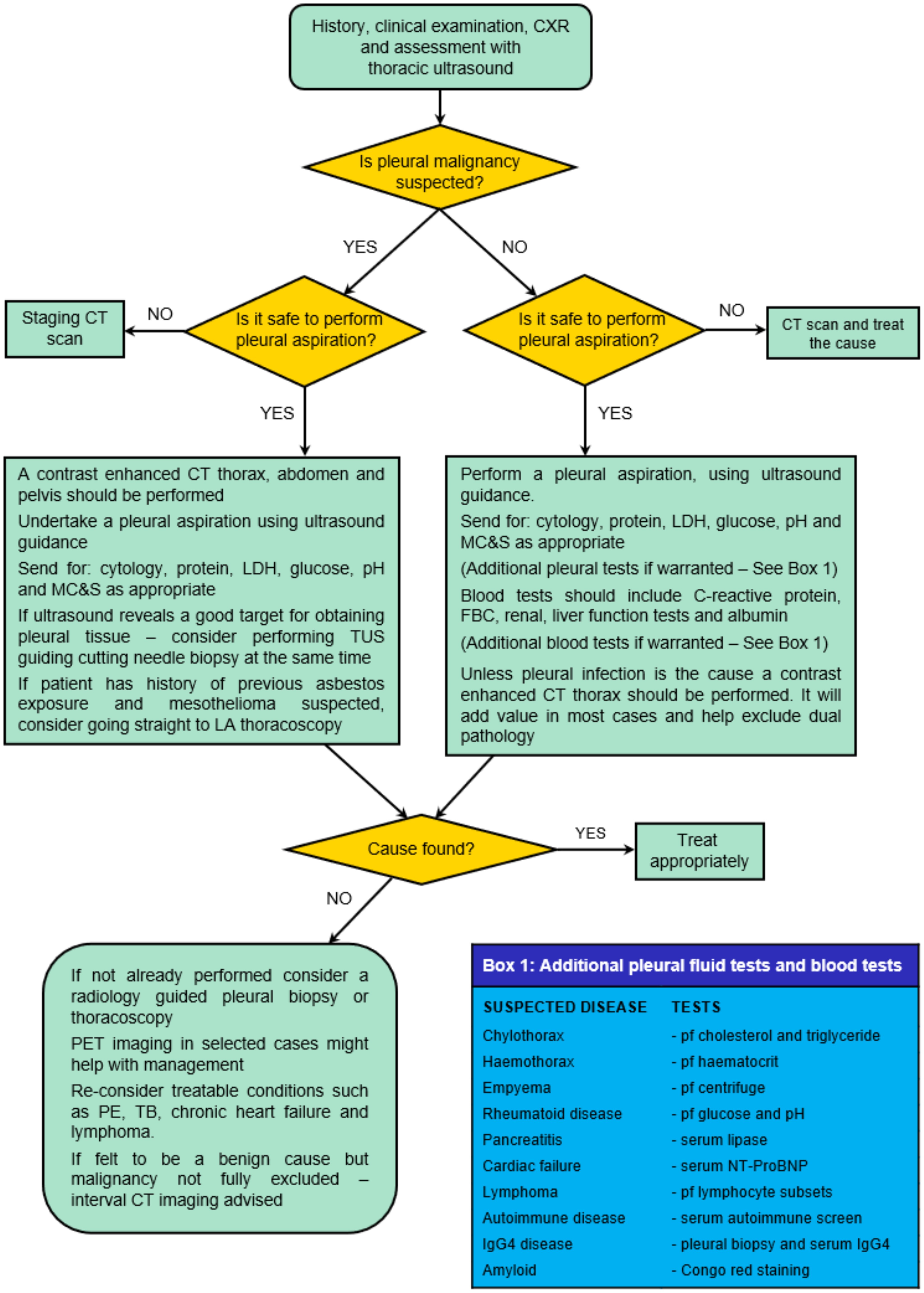
What is the diagnostic accuracy of serum biomarkers?

Unilateral pleural effusion may result from a variety of conditions, including malignant, inflammatory, infectious and cardiovascular illnesses. Serum biomarkers that directly reflect underlying pathophysiology have the potential to shorten diagnostic pathways, either by obviating the need for invasive pleural investigations or by directing interventions such as tissue biopsy or fluid drainage. As for the review on pleural fluid tests (‘What is the diagnostic accuracy of pleural fluid tests (biomarkers)?’ section above), it was again necessary to define the disease states that are of clinical interest in adults presenting with unilateral effusion and to define a relevant gold standard for each (please see table 9 for details), as the index tests reviewed varied with target disease. The next clinical question was:

  • B5 What is the diagnostic accuracy of serum biomarkers when diagnosing adult patients with unilateral pleural effusion?

A summary of the evidence review for each disease state (table 9) is shown in the ‘Secondary pleural malignancy’, ‘Tuberculous pleural effusion’, ‘Heart failure’ and ‘Pleural infection (CPPE or empyema) and autoimmune pleuritis’ subsections below. This is followed by evidence statements, recommendation and GPPs and the full evidence review is available in online supplemental appendix B5.

Secondary pleural malignancy

The sensitivity and specificity of serum CA15-3, CEA, C reactive protein (CRP) and CYFRA21-1 for diagnosing secondary pleural malignancy is shown in table 14. Please note that all presented data are based on data from single studies.

Table 14: Summary of the diagnostic accuracies of secondary pleural malignancy serum biomarkers

Biomarker

Studies (n)

      Cut-Point

Sensitivity

Specificity

CRP

1

              35.5mg/L

0.71

0.56

CYFRA 21-1

1

              3.12 ng/ml

0.71

0.93

CEA

1

              3.35mg/L

0.57

0.93

CA15-3

1

              30.86 ng/ml

0.49

0.93

CA15-3-cancer antigen 15-3; CEA - carcinoembryonic antigen; CRP - C-reactive protein; CYDRA 21-1 - fragment of cytokeratin 19


Tuberculous pleural effusion

The diagnostic accuracies of serum biomarkers T-spot and TB antibody for diagnosing tuberculous pleural effusion are shown in table 15. Please note again that the presented data come from single studies.

Table 15: Summary of the diagnostic accuracies of tuberculous pleural effusion serum biomarkers

Biomarker

Studies (n)

TPE Prevalence

Sensitivity [95% CI]

Specificity [95% CI]

T-spot

1

41%

0.93  [0.83, 0.97]

0.69  [0.58, 0.78]

TB antibody

1

68%

0.48  [0.35, 0.61]

0.76  [0.55, 0.89]


Heart failure

The diagnostic accuracy of NT-proBNP as a diagnostic serum biomarker for diagnosing heart failure in patients with unilateral pleural effusion is shown in table 16.

Table 16: Summary of the diagnostic accuracy of heart failure pleural effusion serum biomarkers

Biomarker

Contributing studies (n)

Sensitivity [95% CI]

Specificity [95% CI]

NT-proBNP

4

0.90  [0.84, 0.94]

0.88  [0.71, 0.96]

NT-proBNP, N-terminal prohormone brain natriuretic peptide.


Pleural infection (CPPE or empyema) and autoimmune pleuritis

No studies directly reported on the diagnostic accuracy of serum biomarkers to diagnose CPPE, empyema or autoimmune pleuritis in patients with unilateral pleural effusion.

Evidence statements

  • Serum NT-proBNP provides high sensitivity and specificity for diagnosing heart failure in patients with unilateral pleural effusion. (Low)

  • There is insufficient evidence to support the use of serum biomarkers to diagnose secondary pleural malignancy, pleural infection, tuberculous pleural effusion or autoimmune pleuritis in patients with unilateral pleural effusion.

Recommendation

  • Serum NT-proBNP should be considered to support a diagnosis of heart failure in patients with unilateral pleural effusion suspected of having heart failure. (Conditional)

Good practice points

  • Serum biomarkers should not currently be used to diagnose secondary pleural malignancy, pleural infection or autoimmune pleuritis.

  • Serum biomarkers should not routinely be used to diagnose tuberculous pleural effusion, but may be considered in high prevalence areas.

  • Serum biomarkers, including NT-proBNP, should not be used in isolation for diagnosing unilateral pleural effusion as multiple conditions may co-exist.

What is the diagnostic accuracy of pleural biopsy?

Obtaining pleural tissue is often necessary to achieve definitive diagnosis in patients presenting with pleural effusion and/or thickening. There are a variety of pleural biopsy techniques (see the BTS Clinical Statement on Pleural Procedures for further details (1) and the aim of the final clinical question in this section was to assess which biopsy method(s) is/are best for achieving accurate histological diagnosis:

  • B6 What is the diagnostic accuracy of pleural biopsy in adults with suspected pleural disease?

Large heterogeneity in study methodology and result reporting made meta-analysis impossible, so a pragmatic approach was adopted to achieve a structured stepwise narrative approach, focusing on studies where direct comparative data were available. Confirming a diagnosis of malignant pleural disease or pleural infection, specifically tuberculous pleuritis, were both considered. Making a histological diagnosis of non-specific pleuritis (also referred to as other terms such as fibrinous pleurisy and pleural fibrosis) was also considered a genuine and clinically relevant finding when followed-up for at least 12 months.

A summary of the evidence review is shown in table 17 using the definitions shown in box 1.

Box 1: Diagnostic test definition


Definitive diagnosis

A final diagnosis made after getting the results of tests, such as blood tests or biopsies[39]


Diagnostic yield

The likelihood that a test or procedure will provide the information needed to establish a diagnosis[40]


Sensitivity

The conditional probability that a person having a disease will be correctly identified by a clinical test[40]


Specificity

The probability that a person not having a disease will be correctly identified by a clinical test[40]


Table 17: Evidence Review summary of ‘What is the diagnostic accuracy of pleural biopsy?’

Comparison

Summary of Evidence review

Medical versus surgical thoracoscopic pleural biopsy

No difference in diagnostic yield, sensitivity or specificity*

Medical rigid versus medical semi-rigid thoracoscopic pleural biopsy

No difference in ‘intention to treat’ or ‘biopsy successfully obtained’ diagnostic yield

Thoracoscopic pleural biopsy versus image-guided closed pleural biopsy  

Definitive diagnosis and diagnostic yield higher with thoracoscopic pleural biopsy (p = 0.04 for both)

Thoracoscopic pleural biopsy versus blind closed pleural biopsy  

Definitive diagnosis and diagnostic yield higher with thoracoscopic pleural biopsy (p = 0.01 and 0.03 respectively)

CT-guided closed pleural biopsy versus ultrasound-guided closed pleural biopsy

No difference in definitive diagnosis

Closed pleural biopsy using core needle versus Abrams needle

Higher diagnostic yield with Abrams needle (p = 0.02)*

Image-guided closed pleural biopsy versus blind closed pleural biopsy

Higher diagnostic yield with image-guided closed pleural biopsy (p = 0.01)


The evidence statements and recommendations are shown below and the full evidence review is available in online supplemental appendix B6.

Evidence statements

  • There is insufficient evidence to determine the diagnostic test performance comparing awake thoracoscopic pleural biopsy and video-assisted thoracoscopic pleural biopsy under general anaesthesia. (Ungraded)

  • There is no difference in diagnostic yield when using rigid thoracoscopy or semi-rigid thoracoscopy to obtain a pleural biopsy. (Low)

  • Definitive diagnosis is more likely with thoracoscopic pleural biopsy when compared with image-guided closed pleural biopsy. (Low)

  • Diagnostic accuracy appears to be higher with thoracoscopic pleural biopsy when compared with image-guided closed pleural biopsy. (Ungraded)

  • Definitive diagnosis is more likely with thoracoscopic pleural biopsy when compared with blind closed pleural biopsy. (Ungraded)

  • Diagnostic yield appears to be higher with thoracoscopic pleural biopsy when compared with blind closed pleural biopsy. (Very low)

  • There is no difference in diagnostic accuracy between CT-guided closed pleural biopsy and ultrasound-guided closed pleural biopsy. (Very low)

  • Image-guided closed pleural biopsy may increase definitive diagnosis and diagnostic accuracy when compared with blind closed pleural biopsy (for malignant disease and tuberculous pleuritis). (Ungraded)

Recommendations

  • Thoracoscopic or image-guided pleural biopsy may be used depending on the clinical indication and local availability of techniques (including need for control of pleural fluid). (Strong)

  • Blind (non-image-guided) pleural biopsies should not be conducted. (Strong—by consensus)

Pleural Infection

Introduction

Pleural infection remains a common medical problem with significant mortality and morbidity despite a better understanding of the aetiology, pathophysiology and recent advances in management approaches. With a combined incidence of over 80 000 cases per annum in the USA and the UK, and an incidence of 11.2 cases per 100 000 population per year in the UK, pleural infection continues to cause a considerable burden to health systems.(40)

A number of studies have been published demonstrating that the incidence of pleural infection is increasing across the Western world,(41–43) including recent data from the UK,(44) and the precise cause of the increase in infection rate, especially in the elderly population, is as yet unclear.

This guideline is intended to address key areas of new evidence since publication of the last BTS Guideline in 2010,(7) which included the specific following questions addressing adults with pleural infection:

  • What is the best predictor of clinical outcomes? (Question C1)

  • Do pleural fluid or radiology parameters accurately determine which patients should be treated with ICD? (Question C2)

  • What initial drainage strategy provides the best clinical outcomes? (Question C3)

  • Does intrapleural therapy improve outcomes compared with other treatment options (eg, drainage alone or surgical intervention)? (Question C4)

  • Which surgical approach provides the best clinical outcomes? (Question C5)

  • Which method of surgery provides the best clinical outcomes? (Question C6)

Areas of clinical importance not covered by the guideline questions are discussed in the ‘Pleural infection, Other areas of clinical importance not covered by the clinical questions’ section.

Other specific areas that have not been covered in this guideline can be referenced from the Management of pleural infection in adults: British Thoracic Society Pleural Disease Guideline 2010, including pathophysiology and the developmental stages of pleural infection.(7)

Definitions and treatment principles of pleural infections

Pleural infection is defined as bacterial entry and replication in the pleural space (45)—the terms ‘complicated’ and ‘uncomplicated’ PPE have been used, but these terms suggest that an associated pneumonia is always a requirement to establish pleural infection which is not the case. The term ‘empyema’ refers to the macroscopic detection of purulent pleural fluid and represents one end of a spectrum of pleural infection.(46) Here, the term ‘pleural infection’ is used to include both empyema and CPPE.

The cornerstones of management of pleural infection are unchanged since the last guideline. These include early identification of cases and accurate diagnosis (covered in the section on approach to the undiagnosed effusion), prompt and suitable antibiotic therapy, nutrition management and deep vein thrombosis prophylaxis and efficient drainage of infected collections (covered in this guideline) via chest tube and adjunctive therapies including intrapleural agents and ultimately surgical management.(7)

What is the best predictor of clinical outcomes?

Clinical outcomes in pleural infection remain poor, with up to 20% of patients dying after an episode of pleural infection over 12 months, and the requirement for surgery in around 15%.(47,48) Understanding which patients are at greater risk of adverse outcomes may allow clinicians to identify means by which their care can be improved to reduce mortality and morbidity, and potentially target invasive treatment to those at highest risk. Hence, the first clinical question in this section asked whether there are baseline clinicoradiological markers that predict clinically important outcomes from pleural infection:

  • C1 For adults with pleural infection, what is the best predictor of clinical outcomes?

The evidence statements and recommendation are presented below; and the full evidence review is presented in online supplemental appendix C1.

Evidence statements

Microbiology parameters

Based on limited evidence:

  • Pleural infection causative organism does not appear to have an effect on predicting mortality rate, hospital length of stay or the need for thoracic surgery in adults with pleural infection. (Ungraded)

  • Healthcare-acquired pleural infection may increase mortality rate and increase hospital length of stay when compared with community-acquired pleural infection in adults. (Ungraded)

Radiological parameters
  • The presence of septation features on ultrasound in adults with pleural infection may be associated with an increased length of hospital stay and increased need for thoracic surgery when compared with non-septated ultrasound features. (Ungraded)

  • The presence of complex septated ultrasound features may be associated with an increased mortality rate, an increased treatment failure rate and an increased length of hospital stay when compared with complex non-septated ultrasound features. (Ungraded)

  • A PPE CT scoring system* may show acceptable discrimination for predicting mortality and/or the need for surgery. (Ungraded)

*Scoring system based on CT radiological features (pleural contrast enhancement, pleural microbubbles, increased attenuation of extrapleural fat and pleural fluid volume >400 mL) for identifying CPPE and defined as a CT score ≥4.(49)

Clinical parameters

  • Higher RAPID scores (Table 18) appear to indicate an increased risk of mortality (Low) (figure 1) and may indicate an increased length of hospital stay. (Ungraded)

  • The Charlson Comorbidity Index Score (CCIS) is associated with an increased risk of mortality with increased CCIS score. (Ungraded)

Table 18: RAPID score*

Parameter

Measure

Score

Renal

Urea (mmol/L)

< 5.0

5.0 – 8.0

> 8.0

0

1

2

Age

< 50 years

50-70 years

> 70 years

0

1

2

Purulence of pleural fluid

Purulent

Non-purulent

0

1

Infection Source

Community acquired

Hospital acquired

0

1

Dietary factor

Albumin (g/L)

> 27.0

< 27.0

0

1

Risk category

Score 0-2

Score 3-4

Score 5-7

Low risk

Medium risk

High risk

*The RAPID score takes into account serum urea level, age, pleural fluid purulence, infection source and serum albumin levels to risk stratify patients into low-risk, medium-risk or high-risk groups.(68)


Figure 1

Comparison of the risk of mortality with low, medium and high RAPID scores (based on the meta-analysis results from online supplemental appendix C1, table C1d and figures C1a–C1f).


Recommendation

  • RAPID scoring should be considered for risk stratifying adults with pleural infection and can be used to inform discussions with patients regarding potential outcome from infection. (Conditional)

Do pleural fluid or radiology parameters accurately determine which patients should be treated with intercostal drainage?

Where bacteria have translocated into the pleural space, ICD is likely to be required to resolve infection, and here this is termed ‘pleural infection’. The presence of macroscopically purulent pleural fluid is termed empyema and diagnostic of pleural infection, and such cases require ICD. In the absence of purulent pleural fluid, there are challenges in determining when best to treat patients with PPE by ICD. Various parameters are available at the point of considering a diagnosis of pleural infection to inform initial decision-making regarding ICD and classification as CPPE which requires drainage, or uncomplicated parapneumonic effusion which will resolve without recourse to drainage. Such parameters include pleural fluid biochemistry (pH, lactate dehydrogenase (LDH), glucose) and radiological features (TUS and CT). Prompt identification of patients with pleural infection who require drainage is necessary to improve patient outcomes by potentially preventing progression to more advanced stages of empyema, so the next clinical question asked:

  • C2 For adults with pleural infection, do pleural fluid or radiology parameters accurately determine which patients should be treated with intercostal drainage?

The evidence statements, recommendations and GPPs are presented below; and the full evidence review is presented in online supplemental appendix C2.

Evidence statements

  • Pleural fluid pH appears to have a high specificity and high sensitivity for identifying patients who will undergo a complicated clinical course (complicated parapneumonic effusion (CPPE)) and thus require intercostal drainage. (Ungraded)

  • In the context of clinically suspected pleural infection:

  • Pleural fluid pH >7.38 appears to indicate a very low risk of CPPE. (Ungraded)

  • Pleural fluid pH ≤7.15 appears to indicate a high risk of CPPE. (Ungraded)

  • Pleural fluid pH between 7.16 and 7.38 appears to indicate a decreasing risk of CPPE/pleural infection with increasing pH, especially with pH >7.22. (Ungraded)

  • Pleural fluid LDH or glucose measurements appear to be less accurate than pH in initial, independent prediction of which patients should be treated with intercostal drainage. (Ungraded)

  • Pleural fluid pH and glucose are highly correlated, and thus where immediate or accurate pH measurement is not possible, an initial glucose of 4.0 mmol/L (in the non-diabetic patient) indicates a moderate-to-high likelihood of CPPE. (Ungraded)

  • CT pleural fluid contrast enhancement may improve detection of CPPE. (Ungraded)

Recommendations

  • For patients with PPE or suspected pleural infection, where diagnostic aspiration does not yield frank pus, immediate pH analysis should be performed. (Strong—by consensus)

  • For patients with suspected CPPE:

    • If pleural fluid pH is ≤7.2, this implies a high risk of CPPE or pleural infection and an ICD should be inserted if the volume of accessible pleural fluid on ultrasound makes it safe to do so. (Strong—by consensus)

    • If pleural fluid pH is >7.2 and <7.4, this implies an intermediate risk of CPPE or pleural infection. Pleural fluid LDH should be measured and if >900 IU/L ICD should be considered, especially if other clinical parameters support CPPE (specifically ongoing temperature, high pleural fluid volume, low pleural fluid glucose (72 mg/dL ≤4.0 mmol/L), pleural contrast enhancement on CT or septation on ultrasound. (Strong—by consensus)

    • If pleural fluid pH is ≥7.4, this implies a low risk of CPPE or pleural infection and there is no indication for immediate drainage. (Strong—by consensus)

  • In the absence of readily available immediate pleural fluid pH measurement, an initial pleural fluid glucose <3.3 mmol/L may be used as an indicator of high probability of CPPE/pleural infection and can be used to inform decision to insert ICD in the appropriate clinical context. (Strong—by consensus)

Good practice points

  • Clinicians should be mindful of alternative diagnoses that can mimic PPE with a low pH and potential for loculations (eg, rheumatoid effusion, effusions due to advanced malignancy/mesothelioma).

  • Pleural fluid samples taken for pH measurement should not be contaminated with local anaesthetic or heparin (eg, by extruding all heparin from an arterial blood gas syringe) as this lowers pleural fluid pH. Delays in obtaining a pleural fluid pH will also increase pleural fluid pH.

  • In patients where a clinical decision is made not to insert an ICD at initial diagnostic aspiration, regular clinical reviews should be performed and repeat thoracocentesis considered to ensure that CPPE is not missed.

The data derived from this question review have been integrated into an updated decision-making algorithm for the diagnosis of patients with pleural infection which includes both pleural fluid and radiological parameters—see Appendix 1, Suspected pleural infection, non-purulent fluid—initial decision tree.

What initial drainage strategy provides the best clinical outcomes?

Adequate drainage of infected fluid from the pleural space in order to achieve source control is a cornerstone of pleural infection management. There are a number of means by which the infected pleural fluid may be removed, ranging from simple percutaneous aspiration or drainage via chest tube to more invasive thoracoscopic and surgical measures. The next clinical question was:

  • C3 For adults with established pleural infection, what initial drainage strategy provides the best clinical outcomes and includes discussion on initial size of chest tube to be used for the treatment of pleural infection and whether initial surgical management should be considered?

A summary of the evidence review is shown in Table 19 and the evidence statements, recommendation and GPPs are presented below. The full evidence review is presented in online supplemental appendix C3.

Table 19: Evidence Review summary for ‘For adults with established pleural infection, what initial drainage strategy provides the best clinical outcomes?’

Clinical Outcome

Summary of Evidence review (surgical drainage* versus chest tube drainage)

Length of hospital stay

Shorter following surgical drainage †

Need for repeat intervention

Increased with chest tube drainage †

Need for thoracic surgery

Not enough evidence

Patient symptoms

Not reported

Complications

Not enough evidence

Quality of life

Not reported

Mortality

Not enough evidence

*Via video-assisted thoracoscopy surgery or open thoracotomy.

†Meta-analysis not possible, data reported in different formats.

Evidence statements

  • Chest tube bore size appears to have no effect on mortality rate, the need for post-treatment thoracic surgery or the length of hospital stay following chest tube drainage to treat pleural infection in adults, but bore size >14 F may increase post-treatment pain. (Ungraded)

  • Drainage under VATS or open thoracotomy appears to reduce the need for repeat intervention and the length of hospital stay when compared with standard chest tube drainage for the treatment of pleural infection in adults. (Ungraded)

Recommendation

  • Initial drainage of pleural infection should be undertaken using a small bore chest tube (14 F or smaller). (Conditional—by consensus)

Good practice points

  • Due to the lack of supporting evidence, early surgical drainage under VATS or thoracotomy should not be considered over chest tube (‘medical’) drainage for the initial treatment of pleural infection.

  • Due to lack of supporting evidence, medical thoracoscopy should not be considered as initial treatment for pleural infection.

Conclusions from the evidence review (please see above and online supplemental appendix B3) have been integrated into the pleural infection treatment algorithm (see Appendix 1, Pleural infection treatment pathway).

Does intrapleural therapy improve outcomes compared with other treatment options (eg, drainage alone or surgical intervention)?

Pleural infection results in the arrangement to the usual fibrinolytic characteristics of the pleural space and it is well established that fibrin deposition and septations occur during the development of pleural infection. On this basis, intrapleural treatments have been used for many years in an attempt to reduce septations and improve drainage, thereby attempting to avoid the need for more invasive surgical management in patients with pleural infection. Intrapleural therapies include fibrinolytics, combined intrapleural enzyme therapy, saline irrigation and intrapleural antibiotics.

The next question investigated if intrapleural therapies improve clinical outcomes in adults with pleural infection (including tuberculous empyema) compared with other treatment options such as drainage alone or surgical intervention:

  • C4 For adults with pleural infection, does intrapleural therapy improve outcomes compared with other treatment options (eg, drainage alone or surgical intervention)?

A summary of the evidence review is shown in Table 20 and the evidence statements, recommendations and GPPs are presented below. The full evidence review is presented in online supplemental appendix C4.

Table 20: Evidence Review summary for ‘For adults with pleural infection, does intrapleural therapy improve outcomes compared to other treatment options (e.g. drainage alone or surgical intervention)?’

Clinical outcomes

Summary of Evidence review (fibrinolytic treatment versus standard care*) [95% CI]

(Fibrinolytic Treatment)

Streptokinase

Urokinase

TPA plus DNAse

TPA

DNAse

Saline irrigation

Length of hospital stay

No difference

3.9 days shorter [5.9 to 13.7] with urokinase

Shorter with TPA plus DNAse

No difference†

No difference†

No difference†

Need for repeat intervention

No difference†

Not reported

Not enough evidence

Not reported

Not reported

Not reported

Need for thoracic surgery

No difference

Reduced need with urokinase (230/1000 [123 to 435]) compared with 512/1000

Reduced need with TPA plus DNAse†

Reduced need with TPA†

No difference†

Reduced need with saline irrigation†

Patient symptoms‡

Reduced symptoms with streptokinase†

Defervesence achieved 4.2 days faster [0.4-7.9] with urokinase

Reduced symptoms with TPA plus DNAse†

No difference†

No difference†

No difference†

Complications§

Increased with streptokinase (114/1000 [64 to 205] compared with 46/1000)

Not reported

Inconclusive results

Inconclusive results

Inconclusive results

No difference†

Quality of life

Not reported

Not reported

Not reported

Not reported

Not reported

Not reported

Mortality

No difference

Not reported

No difference†

No difference†

No difference†

No difference†

Radiological opacification

Inconclusive results

Increased resolution with urokinase†

Increased resolution with TPA plus DNAse†

No difference†

No difference†

Increased resolution with saline irrigation†

Radiographic resolution of effusion

No difference

Greater resolution with urokinase†

Greater resolution with TPA plus DNAse†

Greater resolution with TPA†

Not reported

Not reported

Pleural thickening

No difference†

Potential reduced pleural thickening with urokinase†

Not reported

Not reported

Not reported

Not reported

Standard care—chest drainage alone or chest drainage with intrapleural placebo.

†Reported in a single study.

‡Including persistent chest pain, cough, fever, breathlessness and debilitation.

§Including chest pain, bleeding, fever and tube blockage/dislodgement.

¶Meta-analysis results reported as per 1000 patients.

TPA, tissue plasminogen activator.

Evidence statements

  • Streptokinase appears to have no effect on mortality (Very low), length of hospital stay (Very low), the need for thoracic surgery (Very low) or radiographic resolution of effusion (Very low) for the treatment of pleural infection.

  • Streptokinase increases post-treatment complications (Very low) when compared with chest drainage alone or placebo for the treatment of pleural infection.

  • Urokinase appears to reduce the need for thoracic surgery (Low), hasten the time to resolution of fever (Very low) and reduce the length of hospital stay (Low) compared with placebo or standard care in adults with pleural infection.

  • TPA plus DNAse appears to reduce the length of hospital stay (Ungraded), reduce the likelihood of persistent fevers (Ungraded) and increase improvements in CXR opacification (Ungraded), when compared with placebo in the treatment pleural infection, but TPA plus DNAse may increase the risk of post-treatment complications (serious and non-serious). (Ungraded)

  • Single agent TPA or single agent DNAse do not appear to improve clinical outcomes when compared with placebo for treating pleural infection. (Ungraded)

  • Saline irrigation (250 mL saline three times a day) may reduce the need for thoracic surgery (Ungraded) but appears to have no impact on mortality (Ungraded), length of hospital stay (Ungraded) or time to resolution of fever (Ungraded) when compared with saline flushes.

Recommendations

  • Combination tissue plasminogen activator (TPA) and DNAse should be considered for the treatment of pleural infection, where initial chest tube drainage has ceased and leaves a residual pleural collection. (Conditional—by consensus)

  • Saline irrigation can be considered for the treatment of pleural infection when intrapleural TPA and DNase therapy or surgery is not suitable. (Conditional—by consensus)

  • Single agent TPA or DNAse should not be considered for treatment of pleural infection. (Conditional—by consensus)

  • Streptokinase should not be considered for treatment of pleural infection. (Conditional)

Good practice points

  • Patient consent should be taken when using TPA and DNase as there is a potential risk of bleeding.

  • When administering TPA plus DNase, the regime of should be 10 mg TPA twice daily (10 mg two times per day)+5 mg DNase two times per day for 3 days, based on randomised controlled trial data. Based on retrospective case series data, 5 mg TPA two times per day+5 mg DNase two times per day for 3 days may be as effective, and can be used if considered necessary.

  • Reduced doses of TPA may be considered in those with a potentially higher bleeding risk (eg, those on therapeutic anticoagulation which cannot be temporarily ceased).

  • For details on administration of intrapleural treatments, please refer to the BTS Clinical Statement on Pleural Procedures.(1)

Conclusions from the evidence review (please see above and online supplemental appendix C4) have been integrated into the pleural infection treatment algorithm (see Appendix 1, Pleural infection treatment pathway).

Which surgical approach provides the best clinical outcomes?

A significant proportion of patients with pleural infection fail to improve following optimal medical therapy, and surgical intervention is then required, accepting that not all patients are suitable to undergo surgical treatment. Precise criteria of when to refer for surgery, or what parameters constitute ‘failed medical therapy’ remain unclear.

Different surgical approaches can be used to access the infected space in pleural infection; and these are broadly classified as endoscopic techniques, termed VATS, or open techniques, termed thoracotomy. The next clinical question assessed what the optimal surgical approach is for treating patients with pleural infection:

  • C5 For adults with pleural infection, which surgical approach provides the best clinical outcomes?

A summary of the evidence review is shown in Table 21 and the evidence statements, recommendation and GPP are presented below. The full evidence review is presented in online supplemental appendix C5.

Table 21: Evidence Review summary for ‘Which surgical approach provides the best clinical outcomes?’

Clinical Outcome

Summary of Evidence review (VATS versus thoracotomy) [95% CI]

Length of hospital stay

2.3 days shorter [1.2 to 3.4] with VATS†

Need for repeat intervention

No difference and very low for both (≈ 35/1000)

Patient symptoms

Less post-operative pain with VATS†

Complications

Less with VATS (152/1000 patients [138 to 167] compared with 197/1000 *

Quality of life

Not reported in any study

Mortality

Slightly lower with VATS (35/1000 [29 to 42]) compared with 47/1000 *

*Meta-analysis results reported as per 1000 patients.

†Meta-analysis not possible, data reported in different formats.

VATS, video-assisted thoracoscopy surgery.

Evidence statements

  • Postoperative mortality and the need for repeat intervention are similar following VATS or thoracotomy for pleural infection. (Very low)

  • Immediate postoperative pain appears to be less following VATS than thoracotomy for pleural infection. (Ungraded)

  • Length of hospital stay appears to be shorter following VATS than thoracotomy for pleural infection. (Very low)

  • VATS access appears to cause fewer postoperative complications than thoracotomy for pleural infection. (Very low)

Recommendation

  • VATS access should be considered over thoracotomy for adults in the surgical management of pleural infection. (Conditional)

Good practice point

  • When selecting a surgical access for the treatment of pleural infection in adults, it is important to ensure the technique can facilitate optimal clearance of infected material and achieve lung re-expansion where appropriate.

Which method of surgery provides the best clinical outcomes?

In parallel to the surgical approaches described in the ‘Which surgical approach provides the best clinical outcomes?’ section above (and corresponding online supplemental appendix C5), at the referral point for surgery, different surgical methods can be deployed, broadly classified into drainage, debridement and visceral decortication. The final clinical question in this section therefore aimed to investigate what the best surgical method is for treating pleural infection:

  • C6 For adults with pleural infection, which method of surgery provides the best clinical outcomes?

The evidence statement and GPPs are presented below; and the full evidence review is presented in online supplemental appendix C6.

Evidence statement

  • Based on very limited evidence, decortication surgery for pleural infection may be associated with a longer postoperative stay and higher mortality than surgery that does not involve decortication, but is associated with less breathlessness. (Ungraded)

Recommendations

No recommendations can be made based on the available evidence.

Good practice points

  • Extent of surgery should be tailored according to patient and empyema stage when the lung is not completely trapped (drainage vs debridement).

  • Decortication should be a decision that is individualised to the patient with a trapped lung based on assessment of patient fitness and empyema stage.

Other areas of clinical importance not covered by the clinical questions

Microbiology

The microbiology of pleural infection is a large topic in itself, and not specifically covered by this guideline. However, knowledge of likely microbiological cause will influence the required antibiotic therapy which in a significant proportion of patients will be empirical throughout the treatment course. Large-scale studies have demonstrated that conventional microbiological tests (culture of pleural fluid in plain tubes) results in a sensitivity of 50%–60% at best, with blood cultures having a yield of <10%. There is now good evidence that microbiological yield can be increased by inoculating pleural fluid into blood culture bottle media, and by the use of image-guided parietal pleural biopsy for microbiological assessment (covered in the ‘Investigation of the undiagnosed unilateral pleural effusion, What is the optimal volume and container for a pleural aspiration sample?’ section of this guideline).

Data on likely microbiological cause in pleural infection are little changed compared with the 2010 Guideline,(7) and it remains the case that the majority of community-acquired pleural infection is caused by Gram-positive aerobic organisms, especially streptococcal species including the anginosus group and Staphylococcus aureus. Gram-negative bacteria are less commonly cultured in community-acquired disease, but anaerobic bacteria are commonly seen both in isolation and as co-infection with aerobic organisms. In contrast, hospital-acquired pleural infection is dominated by resistant Gram-positive organisms (including methicillin-resistant Staphylococcus aureus (MRSA)) and Gram-negative organisms such as Escherichia coli, Enterobacter and Pseudomonas, with significant anaerobic involvement. Polymicrobial infection is commonly seen in both community-acquired and hospital-acquired disease, especially when molecular diagnostic techniques are used.

Fungal pleural infection is rare (<1% of cases overall),(50) and usually seen in immunosuppressed individuals, associated with a very high mortality.(51) The diagnosis of fungal pleural infection (especially in those without known immunocompromise) should prompt investigation for other sources of potential infection including oesophageal leak.

Antibiotics

Initial antibiotic treatment for suspected or confirmed pleural infection should commence before results of culture tests are available and will be dictated by the likely source of infection (community-acquired or hospital-acquired disease) as per the likely microbiological cause. Local audit and assessment of likely bacteria is encouraged, as there are geographic differences in microbiological pattern. Initial treatment should include cover of likely organisms including anaerobes, and discussion with local microbiological services should occur. An example of empirical initial antibiotic choice in community-acquired pleural infection is a combination of a second-generation cephalosprin with anaerobic cover (cefuroxime+metronidazole) which covers all likely organisms, whereas empirical treatment for hospital-acquired pleural infection should cover resistant Gram-negative organisms and potentially MRSA (eg, vancomycin+meropenem). A positive culture test from pleural fluid or blood culture may allow narrowing of empirical antibiotics, specifically if pneumococcus is detected, as this is usually a monomicrobial disease.

In general, between 2 and 6 weeks of antibiotic therapy is used according to clinical response, as shorter courses may result in earlier clinical relapse. However, there has not, to date, been an adequately powered study addressing shorter antibiotic duration for pleural infection, and the optimal length of treatment therefore remains unknown. Similarly, direct comparative studies of the use of intravenous or oral antibiotics for pleural infection are lacking, and it is therefore usual practise to treat with intravenous antibiotics initially while patients are in hospital, transitioning to suitable oral therapy according to clinical response and on discharge from hospital. Where oral therapy is not possible (due to bacterial sensitivities or drug allergies), consideration should be given to ambulatory services providing home intravenous treatment.

Pleural Malignancy

Introduction

MPE are of increasing incidence with around a global incidence of around 70 per 100 000 per year.(52) The most common causes of secondary pleural malignancy are lung cancer and breast cancer. Other common primary sites for pleural metastasis include lymphoma, gastrointestinal malignancy and genitourinary malignancy. Malignant pleural mesothelioma (MPM) is a common cause of MPE but is not specifically addressed as part of this guideline, although principles of fluid management and pleurodesis remain similar. MPM is specifically addressed in the British Thoracic Society Guideline for the investigation and management of pleural mesothelioma 2018.(10) The management of MPE has developed hugely since the publication of the management of an MPE: British Thoracic Society Pleural Disease Guideline 2010.(6) Where previously pleural fluid cytology and chest tubes with talc pleurodesis were the mainstay of diagnosis and treatment, there are now many evidence-based options available to clinicians. Medical thoracoscopy is now more widely available and allows the combination of diagnosis and treatment (please see the ‘Medical thoracoscopy’ section in the BTS Clinical Statement on Pleural Procedures for further details(1)). Ambulatory pathways are now a realistic option for patients, and there is evidence for the relative effectiveness of each option.

Diagnosis, treatment and prognosis

Diagnosis

When a patient presents with MPE breathlessness is a common symptom,(53) although up to a quarter of those presenting may not be breathless.(54) Constitutional symptoms, such as fever, chills, fatigue, weakness and weight loss, may be prominent and other symptoms, such as chest pain, are usually because of malignant infiltration of structures in the chest wall.

MPE can be diagnosed by radiology, pleural aspiration under ultrasound guidance or image-guided pleural biopsy. The diagnostic accuracy of various imaging modalities used for diagnosing MPE are explored in the first clinical question and provide an understanding of the relative merits of each technique (see the ‘Which imaging modality is best for diagnosing adults with suspected pleural malignancy?’ section below).

Diagnostic pleural aspiration under ultrasound guidance also remains an important first intervention and may lead to a diagnosis in many cases. Since the widespread adoption of medical thoracoscopy, diagnosis and management of MPE may be combined. This provides a ‘one-stop’ intervention for patients and may significantly shorten the patient pathway. Image-guided pleural biopsy may also be useful where there is significant volume of disease, but little pleural fluid or limited availability of medical thoracoscopy (please see the ‘Ultasound-guided pleural biopsy’ section in the BTS Clinical Statement on Pleural Procedures for further details(1)). Both of these techniques are discussed in the ‘Investigation of the undiagnosed unilateral pleural effusion, Is image guided intervention better than non-image guided intervention?’ section.

Treatment

For patients with a known MPE, management options now include ambulatory intermittent intervention with recurrent aspiration, home-based management with indwelling pleural catheters (IPCs) (also combined with pleurodesis) and traditional inpatient admission with a chest tube and talc slurry pleurodesis. In this guideline, the relative merits of each approach are explored, but it is important to stress that in most cases there is not a ‘right’ answer as to the best approach. Patient preference is always important and various online tools and documents have been developed to help patients navigate the various options. The presented evidence will allow clinicians and patients to make the right decision, including the relative value of pleurodesis and optimum drainage strategies (see ‘Is pleural aspiration with no pleurodesis agent better than talc slurry?’, ‘Is an indwelling pleural catheter better than talc slurry pleurodesis?’, ‘Is thoracoscopy and talc poudrage pleurodesis better than chest drain and talc slurry pleurodesis?’, ‘Is surgical pleurodesis or surgical decortication better than talc slurry pleurodesis?’, ‘Is symptom-based/conservative drainage better than daily drainage?’ and ‘Do intrapleural agents (talc or other pleurodesis agents) improve clinical outcomes in patients with MPE treated with an indwelling pleural catheter?’ sections below). Please note that all procedural techniques are covered separately in the BTS Clinical Statement on Pleural Procedures.(1)

For patients with complex malignant pleural disease, the management of trapped lung is an important question, and the guideline has addressed this in the ‘Is pleural aspiration, talc slurry pleurodesis, talc poudrage pleurodesis or decortication surgery better than using an indwelling pleural catheter to treat malignant pleural effusion and non-expandable lung?’ section below. The use of fibrinolytics for complex pleural effusions has also been explored in the ‘Is intrapleural chemotherapy better than systemic treatment for treating pleural malignancy?’ section.

Finally, the value of systemic anticancer treatment (SACT) or intrapleural chemotherapy for the treatment of MPE are investigated in the ‘Does systemic therapy avoid the need for definitive pleural intervention?’ and ‘Is intrapleural chemotherapy better than systemic treatment for treating pleural malignancy?’ sections, respectively.

Prognosis

Prognosis is a question that is frequently asked, but one which can be difficult to answer. The final clinical question aimed to address if prognostic or predictive scores could provide patients wih MPE with important information about their prognosis (‘Does the use of prognostic or predictive scores provide important prognostic information for the patient?’, Question D12).

The full list of clinical questions in relation to the management of malignant pleural disease in adults were:

  • Which imaging modality is best for diagnosing adults with suspected pleural malignancy? (Question D1)

  • Does systemic therapy avoid the need for definitive pleural intervention? (Question D2)

  • Is pleural aspiration with no pleurodesis agent better than talc slurry? (Question D3)

  • Is an indwelling pleural catheter better than talc slurry pleurodesis? (Question D4)

  • Is thoracoscopy and talc poudrage pleurodesis better than chest drain and talc slurry pleurodesis? (Question D5)

  • Is surgical pleurodesis or surgical decortication better than talc slurry pleurodesis? (Question D6)

  • Is pleural aspiration, talc slurry pleurodesis, talc poudrage pleurodesis or decortication surgery better than using an indwelling pleural catheter to treat malignant pleural effusion and non-expandable lung? (Question D7)

  • Are intrapleural enzymes better than surgery, or no treatment, for treating malignant pleural effusion and septated effusion (on radiology)? (Question D8)

  • Is symptom-based/conservative drainage better than daily drainage? (Question D9)

  • Do intrapleural agents (talc or other pleurodesis agents) improve clinical outcomes in patients with MPE treated with an indwelling pleural catheter? (Question 10)

  • Is intrapleural chemotherapy better than systemic treatment for treating pleural malignancy? (Question D11)

  • Does the use of prognostic or predictive scores provide important prognostic information for the patient? (Question D12)

Topics covered

Topics not covered in the ‘Pleural malignancy’ section

The following topics have not been covered in the ‘Pleural malignancy’ section:

  1. The management of malignant mesothelioma as this is covered in the BTS Guideline for the investigation and management of pleural mesothelioma.(10)

  2. The size of chest tube for optimum drainage, as recent data adequately address this.(55)

  3. The importance of maintaining tube patency and securing the drain to prevent dislodgement cannot be overemphasised but have not been specifically covered in this guideline.

  4. Patient rotation is no longer common practice and hence has not been specifically addressed.

  5. Tube clamping and removal, while important, are addressed indirectly by studies addressing pleurodesis agents (see ‘Is pleural aspiration with no pleurodesis agent better than talc slurry?’, ‘Is an indwelling pleural catheter better than talc slurry pleurodesis?, ‘Is thoracoscopy and talc poudrage pleurodesis better than chest drain and talc slurry pleurodesis?’ and ‘Is surgical pleurodesis or surgical decortication better than talc slurry pleurodesis?’ sections).

  6. Malignant tract seeding is a problem that frequently arises in the management of malignant mesothelioma, but less often in other pleural malignancies, therefore has not been covered within this guideline.

Topics of important consideration

Although there are clinical questions that have not been addressed, patient safety is an important issue that must be considered. While this has not been the subject of rigorous investigative methodology, rate of drainage of an MPE should be addressed. In the UK, various alerts from the National Patient Safety Agency have appeared over recent years (https://www.england.nhs.uk/patient-safety/national-patient-safety-alerting-committee/). Consensus would suggest that the maximum rate of drainage for an MPE in a closed system would be 1.5 L in the first hour, with an hourly rate of 1 L thereafter until drainage is complete. More rapid drainage can be associated with lung re-expansion that is too rapid and the phenomenon of re-expansion pulmonary oedema, which carries significant morbidity and mortality (see the ‘Pleural aspiration (diagnostic and therapeutic), Complications’ section in the BTS Clinical Statement on Pleural Procedures for further details(1)).

Which imaging modality is best for diagnosing adults with suspected pleural malignancy?

Detailed radiological evaluation is commonly performed as part of the assessment for patients with clinical or X-ray findings raising the possibility of pleural malignancy. A range of imaging tools are available including TUS, CT, PET-CT and MRI. Histological confirmation is the gold standard for diagnosis of pleural malignancy, but where tissue sampling is not possible a clinical diagnosis may be made on the basis of disease behaviour over time. Hence, the first clinical question in this section evaluated the diagnostic accuracy of radiological tests to distinguish malignant from benign pleural pathology:

  • D1 What is the diagnostic accuracy of radiology in adults with suspected pleural malignancy?

A summary of the diagnostic accuracies of TUS, CT and PET-CT is shown in table 22 (taken from online supplemental appendix D1, Table D1e).

Table 22: Summary of the diagnostic accuracy of thoracic ultrasound (TUS), CT and PET-CT


Diagnostic accuracy


Modality

Pooled Sensitivity [95% CI]

Pooled Specificity [95% CI]

No. studies

TUS*

0.80 [0.70, 0.87]

0.90 [0.81, 0.94]

2

CT

0.80 [0.62, 0.90]

0.81 [0.72, 0.88]

6

PET-CT

0.89 [0.80, 0.95]

0.92 [0.88, 0.95]

2

*Studies performed in patients with pleural effusion suspected of pleural malignancy.


The use of MRI for evaluation of the pleura is relatively uncommon, but three studies, each assessing different techniques, met the criteria for inclusion. One study assessed the morphological assessment of the pleura, using criteria by Leung et al (56) with MRI, and found it comparable to CT assessment (sensitivity 0.98, specificity 0.92).(57) However, MRI was better able to detect subtle chest wall and/or diaphragmatic infiltration than CT. Malignant pleural disease also tended to be hyperintense on T2-weighted images and gadolinium-enhanced T1-weighted images, unlike benign disease. A second study identified the presence of multiple hyperintense foci on the pleura on high b-value diffusion-weighted images (‘pointillism’) as a marker of malignancy (sensitivity 0.93, specificity 0.79).(58) The final study described a novel marker of pleural malignancy defined by early contrast enhancement on dynamic contrast-enhanced images which, when combined with recognised morphological features, resulted in a sensitivity and specificity of 0.92 and 0.78, respectively. However, comparison with CT evaluation in the same cohort (sensitivity 0.56, specificity 0.77) did not show a statistically significant difference.(59)

The evidence statements, recommendations and GPPs are presented below, and the full evidence review is available in online supplemental appendix D1.

Please also note that the presented data should be supplemented by reference to Section 5 of the BTS Guideline for the investigation and management of pleural mesothelioma.(10)

Evidence statements

  • Ultrasound allows detailed evaluation of the peripheral pleura in the presence of a pleural effusion and has a moderate sensitivity and high specificity for diagnosing pleural malignancy. (Moderate)

  • CT has a moderate sensitivity and specificity for the diagnosis of pleural malignancy. (Low)

  • PET-CT has a high sensitivity and specificity for the diagnosis of pleural malignancy. (Low)

  • MRI, using different techniques, appears to show high sensitivity and specificity for the diagnosis of pleural malignancy. (Ungraded)

Recommendations

  • Ultrasound may be a useful tool at presentation to support a diagnosis of pleural malignancy, particularly in the context of a pleural effusion, where appropriate sonographic skills are present. (Conditional)

  • CT allows assessment of the entire thorax, and positive findings may support a clinical diagnosis of pleural malignancy when biopsy is not an option (Conditional), however a negative CT does not exclude malignancy. (Strong—by consensus)

  • PET-CT can be considered to support a diagnosis of pleural malignancy in adults when there are suspicious CT or clinical features and negative histological results, or when invasive sampling is not an option. (Conditional)

Good practice points

  • Imaging can play an important role in the assessment of pleural malignancy, but results should be interpreted in the context of clinical, histological and biochemical markers.

  • Features of malignancy may not be present on imaging at presentation. Unless a clear diagnosis is reached by other means (eg, biopsy), monitoring with follow-up imaging of patients presenting with pleural thickening and unexplained unilateral pleural effusion should be considered to exclude occult malignancy.

  • MRI has potential as a diagnostic tool in pleural malignancy. Its clinical value has yet to be determined, and its use should be limited to highly selected cases and research studies at the present time.

Does systemic therapy avoid the need for definitive pleural intervention?

MPE often recur after initial aspiration.(6) Since MPE is a marker of advanced disease and is associated with a poor prognosis, treatment focuses on palliation of symptoms and maintenance of quality of life. Anecdotal reports suggest that MPEs often resolve rapidly after initiation of chemotherapy, avoiding the need for a definitive procedure, so the next clinical question aimed to determine whether SACT reduces the requirement for pleural drainage and pleurodesis, with specific focus on treatment-sensitive tumours:

  • D2 For adults with malignant pleural effusion, does systemic therapy avoid the need for definitive pleural intervention?

The evidence statements, recommendation and GPPs are presented below and the full evidence review is available in online supplemental appendix D2.

Evidence statements

  • There was no evidence to support the use of SACT to reduce the need for definitive pleural procedures in adults with MPE.

  • Systemic anti-angiogenesis agents may improve pleural effusion control in non-small-cell lung carcinoma, but methodological constraints limit the interpretation of the results.

Recommendation

  • Definitive pleural intervention should not be deferred until after SACT. (Conditional—by consensus)

Is pleural aspiration with no pleurodesis agent better than talc slurry?

Chest drain insertion with talc pleurodesis provides definitive management of MPE by creating permanent fusion of the pleural layers. This requires hospitalisation with a chest drain in situ for a number of days. Pleural aspiration with no attempt at pleurodesis is an alternative approach and has the advantage of not requiring hospital admission but fluid may recur. Understanding which of these interventions has the most benefit for important clinical outcomes would permit rational treatment choices, so the next clinical question was:

  • D3 For adults with malignant pleural effusion, is pleural aspiration with no pleurodesis agent better than talc slurry at improving clinical outcomes?

The evidence statements, recommendation and GPPs are presented below, and the full evidence review is available in online supplemental appendix D3.

Evidence statements

Based on very limited evidence:

  • Talc slurry pleurodesis may be associated with a longer hospital stay than pleural aspiration. (Ungraded)

  • Talc slurry pleurodesis appears to reduce the need for re-intervention and reduces the overall number of complications compared with pleural aspiration alone. (Ungraded)

  • Patients undergoing pleural aspiration as the first intervention will often require a second procedure, with approximately one-third requiring this within 2 weeks. (Ungraded)

  • Pleural aspiration appears to improve breathlessness. (Ungraded)

Recommendation

  • Management of MPE using talc pleurodesis (or another method) is recommended in preference to repeated aspiration especially in those with a better prognosis, but the relative risks and benefits should be discussed with the patient. (Conditional—by consensus)

Good practice points

  • Decisions on the best treatment modality should be based on patient choice.

  • Informed decision-making should include the role of inpatient versus ambulatory management and the potential risk of requiring further pleural interventions.

Is an indwelling pleural catheter better than talc slurry pleurodesis?

Chest drain insertion with talc pleurodesis and IPCs provide definitive treatment options in the management of MPE. Talc pleurodesis has long been considered the standard of care, however, understanding the role of IPCs in comparison is key to provide optimal options to patients with MPE. This led to the next clinical question:

  • D4 For adults with malignant pleural effusion, is an indwelling pleural catheter better than talc slurry pleurodesis at improving clinical outcomes?

A summary of the evidence is shown in Table 23 and the evidence statements, recommendation and GPPs are presented below. The full evidence review is available in online supplemental appendix D4.

Table 23: Evidence Review summary for ‘Is an indwelling pleural catheter better than talc slurry pleurodesis?

Clinical Outcome

Summary of Evidence review (talc slurry pleurodesis versus IPC) [95% CI]

Length of hospital stay

Longer for initial procedure and total inpatient stay with talc slurry*

Pleurodesis rates

Not enough evidence

Need for re-intervention

Increased need with talc slurry (251/1000 compared with 77/1000 [42 to 138]) †

Complications

No difference, but approximately 20-25% of patients are likely to experience complications when treated with either technique 

Symptoms‡

No difference in dyspnoea, but both treatments show significant improvements

Quality of life (QoL)

Improved QoL with talc slurry pleurodesis and IPC, but no significant difference between the two treatments

*Meta-analysis not possible, data reported in different formats.

†Meta-analysis results reported as per 1000 patients.

‡Breathlessness, chest pain.

IPC, indwelling pleural catheter.


Evidence statements

  • Talc slurry pleurodesis and IPCs appear to improve dyspnoea and quality of life scores, but there are no observable differences between the two treatments. (Ungraded)

  • IPC insertion appears to be associated with a shorter length of initial hospital stay at the time of intervention and fewer subsequent inpatient days. (Ungraded)

  • IPCs appear to be associated with a reduced need for further pleural intervention (defined as requirement for a further pleural procedure) when compared with talc slurry pleurodesis. (Moderate)

  • There appears to be no difference in adverse events for patients treated with talc slurry pleurodesis or IPC. (Very low)

Recommendation

  • Patients without known non-expandable lung (for a definition of non-expandable lung please see the ‘Is pleural aspiration, talc slurry pleurodesis, talc poudrage pleurodesis or decortication surgery better than using an indwelling pleural catheter to treat malignant pleural effusion and non-expandable lung?’ section) should be offered a choice of IPC or pleurodesis as first-line intervention in the management of MPE. The relative risks and benefits should be discussed with patients to individualise treatment choice. (Conditional)

Good practice points

  • The psychological implications and potential altered body image aspects of having a semi-permanent tube drain in situ should not be underestimated and must be considered prior to insertion.

  • All patients who have had an IPC inserted should be referred to the community nursing team on discharge for an early assessment of the wound site, symptom control, support with IPC drainage and removal of sutures.

  • Patients and their relatives should be supported to perform community drainage and complete a drainage diary if they feel able to do so, to promote independence and self-management.

  • Complications such as infection refractory to community management, suspected drain fracture, loculations or blockage with persistent breathlessness should be referred back to the primary pleural team for further assessment.

Is thoracoscopy and talc poudrage pleurodesis better than chest drain and talc slurry pleurodesis?

In adults with MPE, talc pleurodesis is commonly used to provide long-term control of fluid. However, there is debate as to the best way to administer talc. This can either be talc slurry (emulsification of talc in normal saline which is then administered via a chest drain) or poudrage (administration of talc powder as an aerosol during thoracoscopy). Both techniques enable effective delivery of talc to the pleural space, but it has been theorised that talc poudrage may allow better coverage of the pleural space as the talc is directly visualised and may be associated with shorter length of stay as talc is delivered at the same procedure as fluid drainage. However, thoracoscopy is a more invasive procedure. The next clinical question assessed if thoracoscopy and talc poudrage is better than chest drain and talc slurry pleurodesis:

  • D5 For adults with MPE is thoracoscopy and talc poudrage pleurodesis better than chest drain and talc slurry pleurodesis at improving clinical outcomes?

A summary of the evidence is shown in Table 24 and the evidence statements, recommendation and GPPs are presented below. The full evidence review is available in online supplemental appendix D5.

Table 24: Evidence Review summary for ‘Is thoracoscopy and talc poudrage pleurodesis better than chest drain and talc slurry pleurodesis?’

Clinical Outcome

Summary of Evidence review (thoracoscopy and talc poudrage pleurodesis versus chest drain and talc slurry pleurodesis) [95% CI]

Length of hospital stay

No difference

Need for re-intervention

Reduced with thoracoscopy and talc poudrage pleurodesis (138/1000 [103 to 189] compared with 206/1000)*

Complications

No difference in the occurrence of one, or more complications, but thoracoscopy and talc poudrage may cause an increased number of complications per patient

Symptoms†

No difference in chest pain or breathlessness

Quality of life (QoL)

No difference

*Meta-analysis results reported as per 1000 patients.

†Breathlessness, chest pain.


Evidence statements

  • There appears to be no difference in health-related quality of life, length of hospital stay, chest pain or breathlessness in adults with MPE treated with chest drain and talc slurry, or thoracoscopy and talc poudrage. (Ungraded)

  • Pleurodesis failure rate may be lower in adults who have thoracoscopy and talc poudrage for the treatment of MPE when compared with chest drain and talc slurry. (Low)

  • There appears to be no difference in the occurrence of one or more complications following treatment with chest drain and talc slurry or thoracoscopy and talc poudrage in adults with MPE (Very low), but thoracoscopy and talc poudrage may cause an increased number of complications per patient. (Ungraded)

Recommendation

  • Talc slurry or talc poudrage may be offered to patients with MPE to control fluid and reduce the need for repeated procedures. (Conditional)

Good practice point

  • Where a diagnostic procedure is being conducted at thoracoscopy (pleural biopsies), if talc pleurodesis is reasonable, this should be conducted during the same procedure via poudrage.

Is surgical pleurodesis or surgical decortication better than talc slurry pleurodesis?

In adults with MPE, talc pleurodesis via slurry or poudrage, IPCs and aspiration are common treatment options and widely available. However, surgical intervention is a treatment option in those able to tolerate surgery, so the next clinical question reviewed if there are relative benefits of using a surgical approach in MPE compared with the above ‘physician’ approach:

  • D6 For adults with malignant pleural effusion, is surgical pleurodesis or surgical decortication better than talc slurry pleurodesis at improving clinical outcomes?

The evidence statements, recommendation and GPPs are presented below, and the full evidence review is available in online supplemental appendix D6.

Evidence statements

There was insufficient evidence to accurately address the question and published evidence was in highly selected, non-randomised patients.

  • Surgical and non-surgical treatments for MPE may improve quality of life and reduce breathlessness. (Ungraded)

  • Surgical MPE treatments may require a longer stay in hospital compared with talc slurry pleurodesis. (Ungraded)

  • VATS with talc pleurodesis may reduce the need for early postsurgery re-intervention. (Ungraded)

  • Pleurodesis failure rates may increase in patients wih MPE with non-expandable lung if thoracoscopic decortication is not performed. (Ungraded)

Recommendation

  • In selected patients deemed fit for surgery, either surgical talc pleurodesis or medical talc slurry can be considered for the management of MPE. The relative risks, benefits and availability of both techniques should be discussed with patients to individualise treatment choice. (Conditional—by consensus)

Good practice points

  • Informed decision-making should include the role of surgery versus ambulatory management with an IPC for the management of MPE in selected patients.

  • Decortication surgery may improve pleurodesis success in patients with MPE with non-expandable lung, but the risks and benefits of IPC and surgical treatment should be discussed with patients, and treatment individualised according to circumstances (eg, fitness to undergo thoracic surgery).

Is pleural aspiration, talc slurry pleurodesis, talc poudrage pleurodesis or decortication surgery better than using an indwelling pleural catheter to treat malignant pleural effusion and non-expandable lung?

Management of patients with non-expandable lung can be challenging. IPCs have become the preferred management technique for these patients, so the next question reviewed the usefulness of using alternative techniques (pleural aspiration, talc slurry pleurodesis, talc poudrage pleurodesis and decortication surgery) to manage non-expandable lung in malignant pleural disease:

  • D7 For adults with malignant pleural effusion and non-expandable lung, is pleural aspiration, talc slurry pleurodesis, talc poudrage pleurodesis or decortication surgery better than using an indwelling pleural catheter at improving clinical outcomes?

There is no well-defined objective definition of what constitutes ‘non-expandable lung’, but for the purposes of this guideline, non-expandable lung has been defined on expert group consensus as radiologically significant (with >25% of the lung not apposed to the chest wall) based on CXR appearances. It should be noted that there is significant interobserver variation in chest radiograph interpretation of the presence of non-expandable lung. Non-expandable lung may be associated with worse prognosis in MPE.(60) Non-expandable lung is preferred as a term to trapped or entrapped lung as it includes both visceral pleural thickening limiting re-expansion and endobronchial obstruction preventing re-expansion.

The evidence statements and GPPs are presented below, and the full evidence review is available in online supplemental appendix D7.

Evidence statements

  • IPCs may improve quality of life and breathlessness in patients with MPE and non-expandable lung but may result in the IPC remaining in situ for a prolonged period (>100 days). IPC carries a small risk of pleural infection in patients with MPE and non-expandable lung. (Ungraded)

  • There is no direct evidence to support the use of talc slurry pleurodesis over IPC, but talc slurry pleurodesis may improve quality of life, symptoms and pleurodesis rate in patients with MPE and <25% lung non-expandable lung. (Ungraded)

  • There is no direct evidence to support the use of talc poudrage pleurodesis over IPC in patients with MPE and non-expandable lung. (Ungraded)

  • Pleurodesis failure rates may increase in patients with MPE and non-expandable lung if thoracoscopic decortication is not performed. (Ungraded)

Recommendations

No recommendations can be made on the use of pleural aspiration, talc slurry pleurodesis, talc poudrage pleurodesis or decortication surgery versus an IPC to control symptoms in patients with MPE and non-expandable lung.

Good practice points

  • Decisions on treatment modality for MPE and non-expandable lung should be based on patient choice, with the relative risks and benefits of each modality discussed with the patient, but patients should be made aware of the limited evidence base regarding treatment options for non-expandable lung.

  • IPCs are effective at controlling symptoms in non-expandable lung and should be considered, but it may be appropriate to undertake pleural aspiration first to assess symptomatic response.

  • Pleural aspiration may result in a need for multiple procedures so alternatives should be discussed with the patient.

  • In patients with radiologically significant (>25%) non-expandable lung requiring intervention for a symptomatic MPE, current evidence suggests the use of an IPC rather than talc pleurodesis.

  • In patients with MPE and <25% non-expandable lung, talc slurry pleurodesis may improve quality of life, chest pain, breathlessness and pleurodesis rates.

  • Decortication surgery may improve pleurodesis success in selected patients with MPE and non-expandable lung, but the risks and benefits of IPC and surgical treatment should be discussed with patients, and treatment individualised according to circumstances (eg, fitness to undergo thoracic surgery).

Are intrapleural enzymes better than surgery, or no treatment, for treating malignant pleural effusion and septated effusion (on radiology)?

Patients with MPE and septated effusion are less likely to benefit from pleural fluid drainage because percutaneous drainage alone cannot effectively drain the effusion. This group of patients has been reported as having a worse prognosis than other patients with MPE.(61) Septated effusions can occur both in inpatients with chest drains and ambulant patients with IPCs. Effective drainage of the pleural space, either by decortication or by intrapleural enzymes, may improve symptoms. However, surgical intervention is invasive, and carries a significant risk of morbidity and mortality, and may not be appropriate in these patients. The next question compares the use intrapleural agents to achieve clearance of septated malignant effusion against surgical intervention or placebo.

  • D8 For adults with malignant pleural effusion and septated effusion (on radiology), are intrapleural enzymes better than surgery, or no treatment at improving clinical outcomes?

No studies compared intrapleural enzymes against surgery; and furthermore, no studies were identified that looked at the role of surgery in patients with septated MPE. One reason for this may be that septated effusion is identified on ultrasound and surgeons have not historically performed ultrasound in their patients. Hence, the lack of literature supports the view that surgery is rarely used for these patients.

A summary of the intrapleural enzymes versus no treatment evidence review is shown in Table 25 and the evidence statements and GPPs are presented below. The full evidence review is available in online supplemental appendix D8.

Table 25: Evidence Review summary for ‘Are intrapleural enzymes better than surgery, or no treatment, for treating malignant pleural effusion and septated effusion (on radiology)?’

Clinical Outcome

Summary of Evidence review (intrapleural enzymes versus no treatment) [95% CI]

Length of hospital stay

Shorter with intrapleural enzymes*

Pleurodesis rates/ Need for re-intervention

Lower with intrapleural enzymes (287/1000 [177 to 464] compared with 377/1000) †

Complications

Not reported

Symptoms‡

Reduced breathlessness with intrapleural enzymes (278/1000 [168 to 470] compared with 480/1000)

Quality of life (QoL)

No difference*

*Based on a single study.
†Meta-analysis results reported as per 1000 patients.
‡Breathlessness, chest pain.

Evidence statements

There was insufficient evidence to determine if intrapleural enzymes are better than surgery at improving clinical outcomes in adults with MPE and septated effusion (on radiology).

For inpatients with a chest drain

  • Intrapleural fibrinolytic treatment may shorten hospital stay in patients with MPE and septated effusion when compared with no treatment. (Ungraded)

  • Intrapleural fibrinolytic treatment appears to decrease pleurodesis failure rate, when compared with no treatment, in patients with MPE and septated effusion. (Very low)

  • Intrapleural fibrinolytic treatment appears to decrease breathlessness, when compared with no treatment, in patients with MPE and septated effusion. (Very low)

For ambulant patients with indwelling pleural catheters

  • Intrapleural fibrinolytics, when compared with no treatment, may improve breathlessness in patients with MPE and septated effusion, but there is a high rate of recurrent symptomatic loculation. (Ungraded)

Recommendations

Due to the lack of supporting evidence, no recommendations can be made on the use of intrapleural enzymes or surgery for treating adults with MPE and septated effusion (on radiology).

Good practice points

  • Intrapleural fibrinolytics can be considered in highly selected symptomatic patients with MPE and septated effusion to try to improve breathlessness.

  • Intrapleural fibrinolytics may be used in patients with MPE and septated effusion and an IPC to improve drainage if flushing the IPC with normal saline or heparinised saline does not improve drainage.

  • Surgery can be considered for palliation of symptoms in a minority of patients with significantly septated MPE and associated symptoms and otherwise good prognosis and performance status.

Is symptom-based/conservative drainage better than daily drainage?

IPCs offer an ambulatory management pathway in patients with refractory MPE. The original studies (TIME2, the second Therapeutic Intervention in Malignant Effusion Trial and AMPLE, the Australasian Malignant Pleural Effusion trial) (62 63) used regimes of alternate day drainages and this has been incorporated in routine practice. There has been interest on the optimal drainage regime, whether a once-daily drainage regime would offer better clinical outcomes than the less frequent standard alternate days or whether it would be better to offer drainage when patients are symptomatic (symptom-based/conservative drainage regimes). Hence, the next question was:

  • D9 For adults with malignant pleural effusion treated with indwelling pleural catheters, does symptom-based/conservative drainage have better clinical outcomes than daily drainage?

A summary of the evidence review is shown in Table 26 and the evidence statements, recommendations and GPPs are presented below. The full evidence review is available in online supplemental appendix D9.

Clinical Outcome

Summary of Evidence review (symptom-based/conservative drainage versus daily drainage) [95% CI]

Length of hospital stay

No difference*

Pleurodesis rates

Lower with symptom-based/conservative drainage (190/1000 [125 to 293] compared with 431/1000) †

Need for re-intervention

Not reported

Complications

No difference

Symptoms‡

No difference

Quality of life (QoL)

Reduced with symptom-based/conservative drainage

*Meta-analysis not possible, data reported in different formats.
†Meta-analysis results reported as per 1000 patients.
‡Breathlessness, chest pain.

Evidence statements

  • Symptoms (breathlessness and chest pain), complications and length of hospital stay appear to be the same for daily drainage, symptom-guided drainage or alternate daily drainage. (Ungraded)

  • There appear to be no differences in the occurrence of complications between daily drainage and symptom-based/conservative drainage regimes. (Low)

  • Daily drainage increases pleurodesis rates when compared with alternate drainage or symptom-based drainage regimes. (Low)

  • Daily drainage may improve quality of life when compared with a symptom-based/conservative drainage approach, but there is no current evidence that daily drainage improves quality of life when compared with alternate daily drainages. (Ungraded)

Recommendations

  • Where IPC removal is a priority, daily IPC drainages are recommended to offer increased rates of pleurodesis when compared with less frequent drainages of symptom-guided or alternate drainage regimes. (Conditional)

  • Patients should be advised that they do not require daily drainage to control symptoms of breathlessness and chest pain if they wish to opt for a less intensive regime. (Strong—by consensus)

Good practice points

  • Decisions on the optimal drainage frequency should be based on patient choice.

  • Informed decision-making should include the explanation of the effect of drainage regimes on the patient-centred outcomes such as breathlessness and the possibility of autopleurodesis during the disease course.

  • Although daily drainage may result in earlier removal of IPC, there may be an associated cost associated with the increased number of drainage events (both to the healthcare system, and to the patient). This has been addressed in a modelling study (2) and should be considered.

Do intrapleural agents (talc or other pleurodesis agents) improve clinical outcomes in patients wih MPE treated with an indwelling pleural catheter?

With the increasing use of IPCs to control breathlessness in patients with MPE, there has been interest in ‘combination’ procedures, where a pleurodesis agent is inserted via a functioning IPC after a period of drainage. The next clinical question assessed the evidence for the clinical benefits of using this strategy:

  • D10 For adults with malignant pleural effusion treated with indwelling pleural catheters, do intrapleural agents (talc or other pleurodesis agents) improve clinical outcomes?

The evidence statements and recommendation are presented below and the full evidence review is available in online supplemental appendix D10.

Evidence statements

Based on one paper:

  • Pleurodesis rates and quality of life may be improved in patients with MPE and expandable lung (defined as >75% of hemithorax) who have talc instilled via an IPC. (Ungraded)

  • Chest pain and breathlessness may be reduced in patients with MPE and expandable lung (defined as >75% of hemithorax) who have talc instilled via an IPC. (Ungraded)

  • Complication rates do not appear to differ between patients with MPE treated with an IPC and talc or placebo. (Ungraded)

Recommendation

  • Instillation of talc via an IPC should be offered to patients with expandable lung where the clinician or patient deems achieving pleurodesis and IPC removal to be important. (Conditional—by consensus)

Is intrapleural chemotherapy better than systemic treatment for treating pleural malignancy?

SACT provides the mainstay of active treatment for all patients with metastatic cancer, including those with disease spread to the pleura. Symptomatic malignant effusions can affect quality of life, breathing and performance status of these patients and hinder their ability to tolerate SACT, with drainage often needed prior to SACT commencing. Historically, some chemotherapy agents were delivered intrapleurally to act as sclerosants to aid pleurodesis. Recently, with the advent of medical thoracoscopy, regular insertion of IPCs and a growing number of novel anticancer treatments including immunological and biological agents, the intent of delivering intrapleural anticancer treatments has expanded beyond obtaining pleurodesis. Hence, the next question investigated if intrapleural anticancer therapies improve clinical outcomes over systemic treatments:

  • D11 For adults with pleural malignancy, is intrapleural chemotherapy better than systemic treatment at improving clinical outcomes?

No studies directly compared intrapleural anticancer therapy with SACT alone, but the five studies included in the review compared:

  1. Intrapleural chemotherapy versus intrapleural combination therapy (chemotherapy plus vascular endothelial growth factor (VEGF) inhibitor);

  2. Intrapleural chemotherapy versus intrapleural sodium chloride;

  3. Intrapleural chemotherapy versus intrapleural combination therapy (chemotherapy plus angiogenesis inhibitor);

  4. Intrapleural chemotherapy;

  5. Intracavitary (mixed intrapleural and intra-abdominal) chemotherapy versus intracavitary combination therapy (chemotherapy plus angiogenesis inhibitor).

The evidence statements, recommendation and GPP are presented below. The full evidence review is available in online supplemental appendix D11.

Evidence statements

There was no direct evidence to support this question; and based on very limited evidence:

  • Intrapleural combination therapies (chemotherapy plus VEGF inhibitor or angiogenesis inhibitor) may improve effusion control and increase quality of life, progression-free survival and survival time when compared with chemotherapy alone.

Recommendation

  • Intrapleural chemotherapy should not be routinely used for the treatment of MPE. (Conditional—by consensus)

Good practice point

  • All patients of good performance status with metastatic malignancy should be considered for SACT as standard of care as per national guidelines.

Does the use of prognostic or predictive scores provide important prognostic information for the patient?

MPE are associated with short survival as, with the exception of MPM, they signify advanced or metastatic disease. Numerous other factors, including patient characteristics, pleural fluid parameters and biochemical and haematological values have been shown to be related to clinical outcomes in MPE, however these findings have often lacked validation in independent cohorts. Relating separate findings to each other, and interpreting them in the context of patients, is also often difficult. By combining prognostic factors into validated scoring systems, these may be more clinically useful, so final clinical question in this section aimed to determine if validated prognostic scores exist for MPE and, if so, their use improves clinical outcomes for adults with MPE (excluding mesothelioma):

  • D12 For adults with pleural malignancy, does the use of prognostic and predictive scores improve clinical outcomes?

No studies compared clinical outcomes in patients who had treatment directed by a prognostic score at baseline compared with those who had treatment directed using standard measures. Two externally validated prognostic scoring systems have been reported for MPE, the LENT (pleural fluid LDH, Eastern Cooperative Oncology Group (ECOG) performance score, neutrophil-to-lymphocyte ratio, tumour type) and PROMISE (pleurodesis response markers in malignant pleural effusion) scores, however the impact of these scores on clinical decision-making and outcomes other than survival has not been evaluated.(64 65)

The LENT score combines pleural fluid LDH levels, ECOG performance status, serum neutrophil-to-lymphocyte ratio (NLR) and underlying tumour type to predict patients at low risk, moderate risk or high risk of mortality.(64) The PROMISE score evaluates seven clinical biomarkers and one pleural fluid biomarker (haemoglobin, CRP, white blood cell count, ECOG performance status, cancer type, pleural fluid tissue inhibitor of metalloproteinases 1 (TIMP1) concentrations and previous chemotherapy or radiotherapy) to predict absolute risk of death at 3 months.(65)

The evidence statements, recommendation and GPPs are presented below. The full evidence review is available in online supplemental appendix D12.

Evidence statements

  • LENT and PROMISE provide estimates of survival for patients with MPE, but neither have been assessed in their ability to improve outcomes. (Ungraded)

Recommendation

No recommendation can be made from the presented evidence.

Good practice points

  • Clinicians may consider using a validated risk score for MPE, if the information is of use in planning treatments or in discussion with patients.

  • Patients with pleural malignancy should be managed in a multidisciplinary way, including referral to specialist palliative care services where appropriate.

References

List of references

  1. British Thoracic Society clinical statement on pleural procedures. Thorax 2023. To be updated.Google Scholar
  2.  Shafiq M , Simkovich S , Hossen S , et al . Indwelling pleural catheter drainage strategy for malignant effusion: a cost-effectiveness analysis. Ann Am Thorac Soc 2020;17:746–53.doi:10.1513/AnnalsATS.201908-615OC pmid:http://www.ncbi.nlm.nih.gov/pubmed/32125880  Google Scholar
  3. Du Rand I , Maskell N . Introduction and methods: British Thoracic Society pleural disease guideline 2010. Thorax 2010;65 Suppl 2:ii1–3.doi:10.1136/thx.2010.137042 pmid: http://www.ncbi.nlm.nih.gov/pubmed/20696689 Google Scholar
  4. Hooper C , Lee YCG , Maskell N , et al . Investigation of a unilateral pleural effusion in adults: British Thoracic Society pleural disease guideline 2010. Thorax 2010;65 Suppl 2:ii4–17.doi:10.1136/thx.2010.136978 pmid:http://www.ncbi.nlm.nih.gov/pubmed/20696692 Google Scholar
  5. MacDuff A , Arnold A , Harvey J , et al . Management of spontaneous pneumothorax: British Thoracic Society pleural disease guideline 2010. Thorax 2010;65 Suppl 2:ii18–31.doi:10.1136/thx.2010.136986 pmid:http://www.ncbi.nlm.nih.gov/pubmed/20696690 Google Scholar
  6. Roberts ME , Neville E , Berrisford RG , et al . Management of a malignant pleural effusion: British Thoracic Society pleural disease guideline 2010. Thorax 2010;65 Suppl 2:ii32–40.doi:10.1136/thx.2010.136994 pmid:http://www.ncbi.nlm.nih.gov/pubmed/20696691 Google Scholar
  7. Davies HE , Davies RJO , Davies CWH , et al . Management of pleural infection in adults: British Thoracic Society pleural disease guideline 2010. Thorax 2010;65 Suppl 2:ii41–53.doi:10.1136/thx.2010.137000 pmid:http://www.ncbi.nlm.nih.gov/pubmed/20696693 Google Scholar
  8. Rahman NM , Ali NJ , Brown G , et al . Local anaesthetic thoracoscopy: British Thoracic Society pleural disease guideline 2010. Thorax 2010;65 Suppl 2:ii54–60.doi:10.1136/thx.2010.137018 pmid:http://www.ncbi.nlm.nih.gov/pubmed/20696694 Google Scholar
  9. Havelock T , Teoh R , Laws D , et al . Pleural procedures and thoracic ultrasound: British Thoracic Society pleural disease guideline 2010. Thorax 2010;65 Suppl 2:i61–76.doi:10.1136/thx.2010.137026 pmid:http://www.ncbi.nlm.nih.gov/pubmed/20696688 Google Scholar
  10. Woolhouse I , Bishop L , Darlison L , et al . British Thoracic Society guideline for the investigation and management of malignant pleural mesothelioma. Thorax 2018;73:i1–30.doi:10.1136/thoraxjnl-2017-211321 pmid:http://www.ncbi.nlm.nih.gov/pubmed/29444986 Google Scholar
  11. Freeman SC , Kerby CR , Patel A , et al . Development of an interactive web-based tool to conduct and interrogate meta-analysis of diagnostic test accuracy studies: MetaDTA. BMC Med Res Methodol 2019;19:81.doi:10.1186/s12874-019-0724-x pmid:http://www.ncbi.nlm.nih.gov/pubmed/30999861 Google Scholar
  12. Guyatt GH , Oxman AD , Vist GE , et al . GRADE: an emerging consensus on rating quality of evidence and strength of recommendations. BMJ 2008;336:924–6.doi:10.1136/bmj.39489.470347.AD pmid:http://www.ncbi.nlm.nih.gov/pubmed/18436948 Google Scholar
  13. Iorio A , Spencer FA , Falavigna M , et al . Use of GRADE for assessment of evidence about prognosis: rating confidence in estimates of event rates in broad categories of patients. BMJ 2015;350:h870.doi:10.1136/bmj.h870 pmid:http://www.ncbi.nlm.nih.gov/pubmed/25775931 Google Scholar
  14. Huguet A , Hayden JA , Stinson J , et al . Judging the quality of evidence in reviews of prognostic factor research: adapting the GRADE framework. Syst Rev 2013;2:71.doi:10.1186/2046-4053-2-71 pmid:http://www.ncbi.nlm.nih.gov/pubmed/24007720  Google Scholar
  15. Bobbio A , Dechartres A , Bouam S , et al . Epidemiology of spontaneous pneumothorax: gender-related differences. Thorax 2015;70:653–8.doi:10.1136/thoraxjnl-2014-206577 pmid:http://www.ncbi.nlm.nih.gov/pubmed/25918121  Google Scholar
  16. Hallifax RJ , Goldacre R , Landray MJ , et al . Trends in the incidence and recurrence of inpatient-treated spontaneous pneumothorax, 1968-2016. JAMA 2018;320:1471–80.doi:10.1001/jama.2018.14299 pmid:http://www.ncbi.nlm.nih.gov/pubmed/30304427 Google Scholar
  17. Brown SGA , Ball EL , Perrin K , et al . Study protocol for a randomised controlled trial of invasive versus conservative management of primary spontaneous pneumothorax. BMJ Open 2016;6:e011826.doi:10.1136/bmjopen-2016-011826 pmid:http://www.ncbi.nlm.nih.gov/pubmed/27625060 Google Scholar
  18. Hallifax R , Laskawiec-Szkonter M , Dobson M , et al . Randomised ambulatory management of primary pneumothorax (RAMPP): protocol of an open-label, randomised controlled trial. BMJ Open Respir Res 2019;6:e000403.doi:10.1136/bmjresp-2019-000403 pmid:http://www.ncbi.nlm.nih.gov/pubmed/31179004  Google Scholar
  19. Chan JWM , Ko FWS , Ng CK , et al . Management of patients admitted with pneumothorax: a multi-centre study of the practice and outcomes in Hong Kong. Hong Kong Med J 2009;15:427–33.pmid:http://www.ncbi.nlm.nih.gov/pubmed/19966346  Google Scholar
  20. Olesen WH , Katballe N , Sindby JE , et al . Surgical treatment versus conventional chest tube drainage in primary spontaneous pneumothorax: a randomized controlled trial. Eur J Cardiothorac Surg 2018;54:113–21.doi:10.1093/ejcts/ezy003 pmid:http://www.ncbi.nlm.nih.gov/pubmed/29509892   Google Scholar
  21. Walker SP , Bibby AC , Halford P , et al . Recurrence rates in primary spontaneous pneumothorax: a systematic review and meta-analysis. Eur Respir J 2018;52:1800864.doi:10.1183/13993003.00864-2018  Google Scholar
  22. Coker RK , Armstrong A , Church AC , et al . BTS clinical statement on air travel for passengers with respiratory disease. Thorax 2022;77:329–50.doi:10.1136/thoraxjnl-2021-218110 pmid:http://www.ncbi.nlm.nih.gov/pubmed/35228307 Google Scholar
  23. British Thoracic Society guidelines on respiratory aspects of fitness for diving. Thorax 2003;58:3–13.doi:10.1136/thorax.58.1.3 pmid:http://www.ncbi.nlm.nih.gov/pubmed/12511710 FREE Full Text Google Scholar
  24. Lal A , Anderson G , Cowen M , et al . Pneumothorax and pregnancy. Chest 2007;132:1044–8.doi:10.1378/chest.06-2719 pmid:http://www.ncbi.nlm.nih.gov/ pubmed/17873199  Google Scholar
  25. Alifano M , Roth T , Broët SC , et al . Catamenial pneumothorax: a prospective study. Chest 2003;124:1004–8.doi:10.1378/chest.124.3.1004 pmid:http://www.ncbi.nlm.nih.gov/pubmed/12970030  Google Scholar
  26. Haga T , Kurihara M , Kataoka H , et al . Clinical-pathological findings of catamenial pneumothorax: comparison between recurrent cases and non-recurrent cases. Ann Thorac Cardiovasc Surg 2014;20:202–6.doi:10.5761/atcs.oa.12.02227 pmid:http://www.ncbi.nlm.nih.gov/pubmed/23698374 Google Scholar
  27. Alifano M . Catamenial pneumothorax. Curr Opin Pulm Med 2010;16:381–6.doi:10.1097/MCP.0b013e32833a9fc2 pmid:http://www.ncbi.nlm.nih.gov/pubmed/20473170 Google Scholar
  28. Flume PA , Strange C , Ye X , et al . Pneumothorax in cystic fibrosis. Chest 2005;128:720–8.doi:10.1378/chest.128.2.720 pmid:http://www.ncbi.nlm.nih.gov/pubmed/16100160  Google Scholar
  29. Spector ML , Stern RC . Pneumothorax in cystic fibrosis: a 26-year experience. Ann Thorac Surg 1989;47:204–7.doi:10.1016/0003-4975(89)90269-5 pmid:http://www.ncbi.nlm.nih.gov/pubmed/2919902  Google Scholar
  30. Tribble CG , Selden RF , Rodgers BM . Talc poudrage in the treatment of spontaneous pneumothoraces in patients with cystic fibrosis. Ann Surg 1986;204:677–80.doi:10.1097/00000658-198612000-00010 pmid:http://www.ncbi.nlm.nih.gov/pubmed/3789838 Google Scholar
  31. Davis PB , di Sant'Agnese PA . Diagnosis and treatment of cystic fibrosis. An update. Chest 1984;85:802–9.doi:10.1016/S0012-3692(16)62421-2 pmid:http://www.ncbi.nlm.nih.gov/pubmed/6373170 Google Scholar
  32. Schuster SR , McLaughlin FJ , Matthews WJ , et al . Management of pneumothorax in cystic fibrosis. J Pediatr Surg 1983;18:492–7.doi:10.1016/S0022-3468(83)80207-3 pmid:http://www.ncbi.nlm.nih.gov/pubmed/6620095 Google Scholar
  33. Penketh AR , Knight RK , Hodson ME , et al . Management of pneumothorax in adults with cystic fibrosis. Thorax 1982;37:850–3.doi:10.1136/thx.37.11.850 pmid:http://www.ncbi.nlm.nih.gov/pubmed/7164004  Google Scholar
  34. Arenas-Jiménez J , Alonso-Charterina S , Sánchez-Payá J , et al . Evaluation of CT findings for diagnosis of pleural effusions. Eur Radiol 2000;10:681–90.doi:10.1007/s003300050984 pmid:http://www.ncbi.nlm.nih.gov/pubmed/10795555  Google Scholar
  35. Yilmaz U , Polat G , Sahin N , et al . CT in differential diagnosis of benign and malignant pleural disease. Monaldi Arch Chest Dis 2005;63:17–22.doi:10.4081/monaldi.2005.653 pmid:http://www.ncbi.nlm.nih.gov/pubmed/16035560   Google Scholar
  36. Hallifax RJ , Haris M , Corcoran JP , et al . Role of CT in assessing pleural malignancy prior to thoracoscopy. Thorax 2015;70:192–3.doi:10.1136/thoraxjnl-2014-206054 pmid:http://www.ncbi.nlm.nih.gov/pubmed/25077699 Google Scholar
  37. Lee HY , Goo JM , Lee HJ , et al . The value of computed tomography for predicting empyema-associated malignancy. J Comput Assist Tomogr 2006;30:453–9.doi:10.1097/00004728-200605000-00017 pmid:http://www.ncbi.nlm.nih.gov/pubmed/16778621  Google Scholar
  38. Bugalho A , Ferreira D , Dias SS , et al . The diagnostic value of transthoracic ultrasonographic features in predicting malignancy in undiagnosed pleural effusions: a prospective observational study. Respiration 2014;87:270–8.doi:10.1159/000357266 pmid:http://www.ncbi.nlm.nih.gov/pubmed/24480900 Google Scholar
  39. Chen H-J , Hsu W-H , Tu C-Y , et al . Sonographic septation in lymphocyte-rich exudative pleural effusions: a useful diagnostic predictor for tuberculosis. J Ultrasound Med 2006;25:857–63.doi:10.7863/jum.2006.25.7.857 pmid:http://www.ncbi.nlm.nih.gov/pubmed/16798896 Google Scholar
  40. Gould J . Pleural infection: a case where clinical improvement was misleading. BMJ Case Rep 2013;2013:bcr2013008700.doi:10.1136/bcr-2013-008700 pmid:http://www.ncbi.nlm.nih.gov/pubmed/23605824  Google Scholar
  41. Farjah F , Symons RG , Krishnadasan B , et al . Management of pleural space infections: a population-based analysis. J Thorac Cardiovasc Surg 2007;133:346–51.doi:10.1016/j.jtcvs.2006.09.038 pmid:http://www.ncbi.nlm.nih.gov/pubmed/17258562  Google Scholar
  42. Grijalva CG , Zhu Y , Nuorti JP , et al . Emergence of parapneumonic empyema in the USA. Thorax 2011;66:663–8.doi:10.1136/thx.2010.156406 pmid:http://www.ncbi.nlm.nih.gov/pubmed/21617169  Google Scholar
  43. Finley C , Clifton J , Fitzgerald JM , et al . Empyema: an increasing concern in Canada. Can Respir J 2008;15:85–9.doi:10.1155/2008/975312 pmid:http://www.ncbi.nlm.nih.gov/pubmed/18354748 Google Scholar
  44. Arnold DT , Hamilton FW , Morris TT , et al . Epidemiology of pleural empyema in English hospitals and the impact of influenza. Eur Respir J 2021;57:2003546.doi:10.1183/13993003.03546-2020 pmid:http://www.ncbi.nlm.nih.gov/pubmed/33334937 Google Scholar
  45. Rosenstengel A . Pleural infection-current diagnosis and management. J Thorac Dis 2012;4:186–93.doi:10.3978/j.issn.2072-1439.2012.01.12 pmid:http://www.ncbi.nlm.nih.gov/pubmed/22833824   Google Scholar
  46. Dyrhovden R , Nygaard RM , Patel R , et al . The bacterial aetiology of pleural empyema. A descriptive and comparative metagenomic study. Clin Microbiol Infect 2019;25:981–6.doi:10.1016/j.cmi.2018.11.030 pmid:http://www.ncbi.nlm.nih.gov/pubmed/30580031   Google Scholar
  47. Ferguson AD , Prescott RJ , Selkon JB , et al . The clinical course and management of thoracic empyema. QJM 1996;89:285–90.doi:10.1093/qjmed/89.4.285 pmid:http://www.ncbi.nlm.nih.gov/pubmed/8733515   Google Scholar
  48. Maskell NA , Batt S , Hedley EL , et al . The bacteriology of pleural infection by genetic and standard methods and its mortality significance. Am J Respir Crit Care Med 2006;174:817–23.doi:10.1164/rccm.200601-074OC pmid:http://www.ncbi.nlm.nih.gov/pubmed/16840746   Google Scholar
  49. Porcel JM , Pardina M , Alemán C , et al . Computed tomography scoring system for discriminating between parapneumonic effusions eventually drained and those cured only with antibiotics. Respirology 2017;22:1199–204.doi:10.1111/resp.13040 pmid:http://www.ncbi.nlm.nih.gov/pubmed/28370693   Google Scholar
  50. Rahman NM , Chapman SJ , Davies RJO . The approach to the patient with a parapneumonic effusion. Clin Chest Med 2006;27:253–66.doi:10.1016/j.ccm.2005.12.005 pmid:http://www.ncbi.nlm.nih.gov/pubmed/16716817  Google Scholar
  51. Ko SC , Chen KY , Hsueh PR , et al . Fungal empyema thoracis: an emerging clinical entity. Chest 2000;117:1672–8.doi:10.1378/chest.117.6.1672 pmid:http://www.ncbi.nlm.nih.gov/pubmed/10858401  Google Scholar
  52. Bodtger U , Hallifax RJ . Epidemiology: why is pleural disease becoming more common? In: Maskell NA , Laursen CB , YCG L , et al. , eds. Pleural Disease (ERS Monograph). Sheffield, European Respiratory Society, 2020: 1–12.Google Scholar
  53. Judson M , Sahn S . Pulmonary physiologic abnormalities caused by pleural disease. Semin Respir Crit Care Med 1995;16:346–53.doi:10.1055/s-2007-1009846  Google Scholar
  54. Chernow B , Sahn SA . Carcinomatous involvement of the pleura: an analysis of 96 patients. Am J Med 1977;63:695–702.doi:10.1016/0002-9343(77)90154-1 pmid:http://www.ncbi.nlm.nih.gov/pubmed/930945 Google Scholar
  55. Rahman NM , Pepperell J , Rehal S , et al . Effect of opioids vs NSAIDs and larger vs smaller chest tube size on pain control and pleurodesis efficacy among patients with malignant pleural effusion: the TIME1 randomized clinical trial. JAMA 2015;314:2641–53.doi:10.1001/jama.2015.16840 pmid:http://www.ncbi.nlm.nih.gov/pubmed/26720026  Google Scholar
  56. Leung AN , Müller NL , Miller RR . CT in differential diagnosis of diffuse pleural disease. AJR Am J Roentgenol 1990;154:487–92.doi:10.2214/ajr.154.3.2106209 pmid:http://www.ncbi.nlm.nih.gov/pubmed/2106209 Google Scholar
  57. Luo L , Hierholzer J , Bittner RC , et al . Magnetic resonance imaging in distinguishing malignant from benign pleural disease. Chin Med J 2001;114:645–9.pmid:http://www.ncbi.nlm.nih.gov/pubmed/11780445  Google Scholar
  58. Coolen J , De Keyzer F , Nafteux P , et al . Malignant pleural mesothelioma: visual assessment by using pleural pointillism at diffusion-weighted MR imaging. Radiology 2015;274:576–84.doi:10.1148/radiol.14132111 pmid:http://www.ncbi.nlm.nih.gov/pubmed/25243538 Google Scholar
  59. Tsim S , Humphreys CA , Cowell GW , et al . Early contrast enhancement: a novel magnetic resonance imaging biomarker of pleural malignancy. Lung Cancer 2018;118:48–56.doi:10.1016/j.lungcan.2018.01.014 pmid:http://www.ncbi.nlm.nih.gov/pubmed/29572002 Google Scholar
  60. Martin GA , Kidd AC , Tsim S , et al . Inter-observer variation in image interpretation and the prognostic importance of non-expansile lung in malignant pleural effusion. Respirology 2020;25:298–304.doi:10.1111/resp.13681 pmid:http://www.ncbi.nlm.nih.gov/pubmed/31433545  Google Scholar
  61. Mishra EK , Clive AO , Wills GH , et al . Randomized controlled trial of urokinase versus placebo for nondraining malignant pleural effusion. Am J Respir Crit Care Med 2018;197:502–8.doi:10.1164/rccm.201704-0809OC pmid:http://www.ncbi.nlm.nih.gov/pubmed/28926296 Google Scholar
  62. Davies H , Mishra EK , Wrightson JM . The second therapeutic intervention in malignant effusion trial (TIME2): a randomised controlled trial to assess the efficacy and safety of patient controlled malignant pleural effusion drainage by indwelling pleural catheter compared to chest tube and talc slurry pleurodesis. Am J Respir Crit Care Med 2012;185:A6861.Google Scholar
  63. Lee YG , Fysh ETH , Thomas R . Australasian malignant pleural effusion (AMPLE) trial: a multicentre randomised study comparing indwelling pleural catheter versus talc pleurodesis. Am J Respir Crit Care Med 2016;193:A7812.Google Scholar
  64. Clive AO , Kahan BC , Hooper CE , et al . Predicting survival in malignant pleural effusion: development and validation of the LENT prognostic score. Thorax 2014;69:1098–104.doi:10.1136/thoraxjnl-2014-205285 pmid:http://www.ncbi.nlm.nih.gov/pubmed/25100651  Google Scholar
  65. Psallidas I , Kanellakis NI , Gerry S , et al . Development and validation of response markers to predict survival and pleurodesis success in patients with malignant pleural effusion (PROMISE): a multicohort analysis. Lancet Oncol 2018;19:930–9.doi:10.1016/S1470-2045(18)30294-8 pmid:http://www.ncbi.nlm.nih.gov/pubmed/29908990 Google Scholar
  66. National Cancer Institute’s Dictionary of Cancer Terms. Available: https://www.cancer.gov/‌publications/‌dictionaries/cancer-terms/.Google Scholar
  67. The Free Dictionary’s Medical dictionary. Available: https://medical-dictionary.thefreedictionary.com/ .Google Scholar
  68. Rahman NM , Kahan BC , Miller RF , et al . A clinical score (RAPID) to identify those at risk for poor outcome at presentation in patients with pleural infection. Chest 2014;145:848–55.doi:10.1378/chest.13-1558 pmid:http://www.ncbi.nlm.nih.gov/pubmed/24264558  Google Scholar

Guideline relevant information

Guideline Group members

The Guideline Development Group (GDG) was chaired by three respiratory consultants—Professor Nick Maskell, Professor Najib Rahman and Dr Mark Roberts. The GDG had a wide membership and included colleagues from respiratory medicine, oncology, radiology, pathology and palliative medicine. Two patient representatives were recruited to the group, but due to personal circumstances both had to withdraw before completion of the guideline (August 2019 and July 2021). However, two further patient representatives were recruited at the end of the guideline process to review the final guideline and provide the patients’ perspective. Those on the group were not required to be BTS members and a full list of members can be seen below.

Dr Mark Roberts (co-chair) Consultant in Respiratory Medicine
Sherwood Forest Hospitals NHS Foundation Trust, Nottinghamshire
Prof Najib Rahman (co-chair) Oxford University Hospitals NHS Foundation Trust, Oxford NIHR Biomedical Research Centre, Oxford Respiratory Trials Unit, University of Oxford
Prof Nick Maskell (co-chair) Professor of Respiratory Medicine, North Bristol NHS Trust
Dr Anna Bibby Consultant in Respiratory Medicine, North Bristol NHS Trust
Prof Kevin Blyth Professor of Respiratory Medicine, Queen Elizabeth University Hospital, Glasgow
Dr John Corcoran Consultant in Respiratory Medicine, University Hospitals Plymouth NHS Trust
Dr Anthony Edey Consultant in Thoracic Radiology, North Bristol NHS Trust
Dr Matthew Evison Consultant in Respiratory Medicine, Manchester University NHS Foundation Trust
Dr Duneesha de Fonseka Consultant in Respiratory Medicine, Sheffield Teaching Hospitals NHS Foundation Trust
Dr Rob Hallifax Consultant in Respiratory Medicine, Oxford University Hospitals NHS Foundation Trust
Dr Susan Harden Consultant in Clinical Oncology, Peter MacCallum Cancer Centre, Melbourne, Victoria, Australia (formerly Cambridge University Hospitals NHS Foundation Trust)
Dr Iain Lawrie Consultant in Palliative Medicine, Manchester University Hospitals NHS Foundation Trust
Prof Eric Lim Consultant in Thoracic Surgery, The Royal Brompton Hospital and Imperial College, London
Dr David McCracken Consultant in Acute and Respiratory Medicine, Belfast Health and Social Care Trust
Dr Rachel Mercer Consultant in Respiratory Medicine, Portsmouth Hospitals University NHS Trust
Dr Eleanor Mishra Consultant in Respiratory Medicine, Norfolk and Norwich University Hospitals NHS Foundation Trust, Norwich
Prof Andrew Nicholson Consultant in Histopathology, The Royal Brompton Hospital and Imperial College, London
Ms Farinaz Noorzad Pleural Practitioner, St George’s University Hospitals NHS Foundation Trust, London
Mrs Maria Parsonage Nurse Consultant in Respiratory Medicine, North Cumbria Integrated Care NHS Foundation Trust
Dr Andrew Stanton Consultant in Respiratory Medicine, Newcastle upon Tyne NHS Foundation Trust
Dr Steven Walker Clinical Lecturer in Respiratory Medicine, North Bristol NHS Trust
Mr Richard Bremner (November 2018 – August 2019)
Mr Yannick Mouchilli Patient representative (November 2018 – August 2020)


Declarations of interests

BTS declarations of interest forms have been completed by all members for each year they were part of the GDG. Details of these forms can be obtained from BTS Head Office. ‘Declarations of interests’ was a standing item at each GDG meeting.

Acknowledgements

The co-chairs would like to acknowledge the huge contributions of all guideline group members both to robust discussions during the meetings and sourcing, critically reviewing papers and formulating judgements. They would also like to specifically thank Dr Kirstie Opstad at BTS Head Office who has coordinated the whole process, performed searches and initial abstract filtering, supported the evidence review process and ensured consistency of presentation of the whole guideline. 

The GDG would like to thank Mr Richard Bremner, Mr Yannick Mouchilli, Mr Chris Smith and Dr Tim Wallington (patient representatives) for their helpful contributions during development of this guideline.

Clinical Questions

Pneumothorax

A1 - Are conservative, aspiration, ambulatory, chemical pleurodesis or surgical interventions better than, or as good as, intercostal drainage at improving clinical outcomes in adult pneumothorax patients?

A2 - In adults who have resolved their first episode of pneumothorax, is surgery a better elective management strategy than non-surgery at improving clinical outcomes?

A3 - In adults with spontaneous pneumothorax and ongoing air leak (excluding post-surgical patients), which treatments are better than ongoing chest tube drainage alone at improving clinical outcomes?

A4 - In adults with spontaneous pneumothorax undergoing surgery, what is the optimal operation for improving clinical outcomes?

A5 - What is the optimal surgical approach when performing pneumothorax surgery?

Investigation of the undiagnosed pleural effusion

B1 - What is the diagnostic accuracy of radiology when diagnosing benign pleural disease as a cause of unilateral pleural effusion in adults?

B2 - For adults with suspected unilateral pleural effusion, is image-guided intervention better than non-image-guided intervention at improving clinical outcomes?

B3 - What is the optimal volume and container for a pleural aspiration sample when diagnosing unilateral pleural effusion in adults?

B4 - What is the diagnostic accuracy of pleural fluid tests when diagnosing adult patients with unilateral pleural effusion?

B5 - What is the diagnostic accuracy of serum biomarkers when diagnosing adult patients with unilateral pleural effusion?

B6 - What is the diagnostic accuracy of pleural biopsy in adults with suspected pleural disease?

Pleural infection

C1 - For adults with pleural infection, what is the best predictor of clinical outcomes?

C2 - For adults with pleural infection, do pleural fluid or radiology parameters accurately determine which patients should be treated with intercostal drainage?

C3 - For adults with established pleural infection, what initial drainage strategy provides the best clinical outcomes?

C4 - For adults with pleural infection, does intrapleural therapy, in comparison to other options (drainage or surgical drainage), improve outcomes?

C5 - For adults with pleural infection, which method of surgery provides the best clinical outcomes?

C6 - For adults with pleural infection, which surgical approach provides the best clinical outcomes?

Pleural Malignancy

D1 - What is the diagnostic accuracy of radiology in adults with suspected pleural malignancy?

D2 - For adults with malignant pleural effusion, does systemic therapy avoid the need for definitive pleural intervention?

D3 - For adults with malignant pleural effusion, is pleural aspiration with no pleurodesis agent better than talc slurry at improving clinical outcomes?

D4 - For adults with malignant pleural effusion, is an indwelling pleural catheter better than talc slurry pleurodesis at improving clinical outcomes?

D5 - For adults with malignant pleural effusion, is thoracoscopy (local anaesthetic or VATS) and talc poudrage pleurodesis better than chest drain and talc slurry pleurodesis at improving clinical outcomes?

D6 - For adults with malignant pleural effusion, is surgery better than talc slurry pleurodesis at improving clinical outcomes?

D7 - For adults with malignant pleural effusion and non-expandable lung, is pleural aspiration, talc slurry pleurodesis, talc poudrage pleurodesis or decortication surgery better than using an indwelling pleural catheter at improving clinical outcomes?

D8 - For adults with malignant pleural effusion and septated effusion (on ultrasound or CT), are intrapleural enzymes better than surgery or no treatment at improving clinical outcomes?

D9 - For adults with malignant pleural effusion treated with indwelling pleural catheters, does symptom-based drainage have better clinical outcomes than daily drainage?

D10 - For adults with malignant pleural effusion treated with indwelling pleural catheters, do intrapleural agents (talc or other pleurodesis agents) improve clinical outcomes?

D11 - For adults with pleural malignancy, is intrapleural chemotherapy better than systemic treatment at improving clinical outcomes?

D12 - For adults with pleural malignancy, does the use of prognostic and predictive scores improve clinical outcomes?

Stakeholder organisations

Stakeholders were identified at the start of the process. All stakeholder organisations were notified when the guideline was available for public consultation and a list is published below.

  • Association for Palliative Medicine of Great Britain & Ireland
  • Association of Respiratory Nurse Specialists
  • British Society of Thoracic Imaging
  • British Thoracic Oncology Group
  • Royal College of Pathologists
  • Royal College of Radiologists
  • Society for Cardiothoracic Surgery in Great Britain & Ireland

Review of the guideline

The guideline will be reviewed 5 years after the date of publication.

Downloadable appendices

Online appendices

Click on the tabs to view the appendices for each section.

No images available.

Download resources

  • Clinical Pathways
  • Research Recommendations
  • Spontaneous Pneumothorax
  • Pleural Effusion
  • Pleural Infection
  • Pleural Malignancy
  • BTS Guideline for Pleural Disease Appendix 1: Clinical Pathways/decision trees
    DOWNLOAD
  • Online Appendix 2 (Pleural Disease): Research recommendations
    DOWNLOAD
  • Online Appendix A1 (Pleural Disease): Pneumothorax best acute management
    DOWNLOAD
  • Online Appendix A2 (Pleural Disease): Pneumothorax first episode resolution optimal management
    DOWNLOAD
  • Online Appendix A3 (Pleural Disease): Pneumothorax ongoing air leak optimal management
    DOWNLOAD
  • Online Appendix A4 (Pleural Disease): Pneumothorax optimal surgical approach
    DOWNLOAD
  • Online Appendix A5 (Pleural Disease): Pneumothorax optimal operation
    DOWNLOAD
  • Online Appendix B1 (Pleural Disease): Pleural effusion radiology diagnostic accuracy
    DOWNLOAD
  • Online Appendix B2 (Pleural Disease): Pleural effusion image guided or non image guided
    DOWNLOAD
  • Online Appendix B3 (Pleural Disease): Pleural effusion optimal volume and container
    DOWNLOAD
  • Online Appendix B4 (Pleural Disease): Pleural effusion pleural fluid test diagnostic accuracy
    DOWNLOAD
  • Online Appendix B5 (Pleural Disease): Pleural effusion serum biomarker diagnostic accuracy
    DOWNLOAD
  • Online Appendix B6 (Pleural Disease): Pleural effusion pleural biopsy diagnostic accuracy
    DOWNLOAD
  • Online Appendix C1 (Pleural Disease): Pleural infection best predictor of clinical outcomes
    DOWNLOAD
  • Online Appendix C2 (Pleural Disease): Pleural infection pleural fluid or radiology parameters
    DOWNLOAD
  • Online Appendix C3 (Pleural Disease): Pleural infection initial drainage strategy
    DOWNLOAD
  • Online Appendix C4 (Pleural Disease): Pleural infection intrapleural therapy
    DOWNLOAD
  • Online Appendix C5 (Pleural Disease): Pleural infection optimal surgical approach
    DOWNLOAD
  • Online Appendix C6 (Pleural Disease): Pleural infection optimal surgical method
    DOWNLOAD
  • Online Appendix D1 (Pleural Disease): Pleural malignancy best imaging modality
    DOWNLOAD
  • Online Appendix D2 (Pleural Disease): Pleural malignancy systemic therapy
    DOWNLOAD
  • Online Appendix D3 (Pleural Disease): Pleural malignancy pleural aspiration versus talc slurry
    DOWNLOAD
  • Online Appendix D4 (Pleural Disease): Pleural malignancy IPC versus talc slurry
    DOWNLOAD
  • Online Appendix D5 (Pleural Disease): Pleural malignancy talc poudrage versus talc slurry
    DOWNLOAD
  • Online Appendix D6 (Pleural Disease): Pleural malignancy surgery versus talc slurry
    DOWNLOAD
  • Online Appendix D7 (Pleural Disease): Pleural malignancy non-expandable lung
    DOWNLOAD
  • Online Appendix D8 (Pleural Disease): Pleural malignancy intrapleural enzymes
    DOWNLOAD
  • Online Appendix D9 (Pleural Disease): Pleural malignancy IPC drainage strategies
    DOWNLOAD
  • Online Appendix D10 (Pleural Disease): Pleural malignancy intrapleural agents
    DOWNLOAD
  • Online Appendix D11 (Pleural Disease): Pleural malignancy intrapleural chemotherapy
    DOWNLOAD
  • Online Appendix D12 (Pleural Disease): Pleural malignancy prognostic scores
    DOWNLOAD